Muscles of the arm in 3D
The muscles of the arm (upper limb) move the shoulder, elbow, wrist and fingers. They are grouped into compartments: flexors at the front of the arm and forearm, extensors at the back, and the intrinsic muscles of the hand. This free 3D model shows the muscles of the right upper limb on its bones and lets you isolate any muscle, view it from every side and remove the layers one by one.
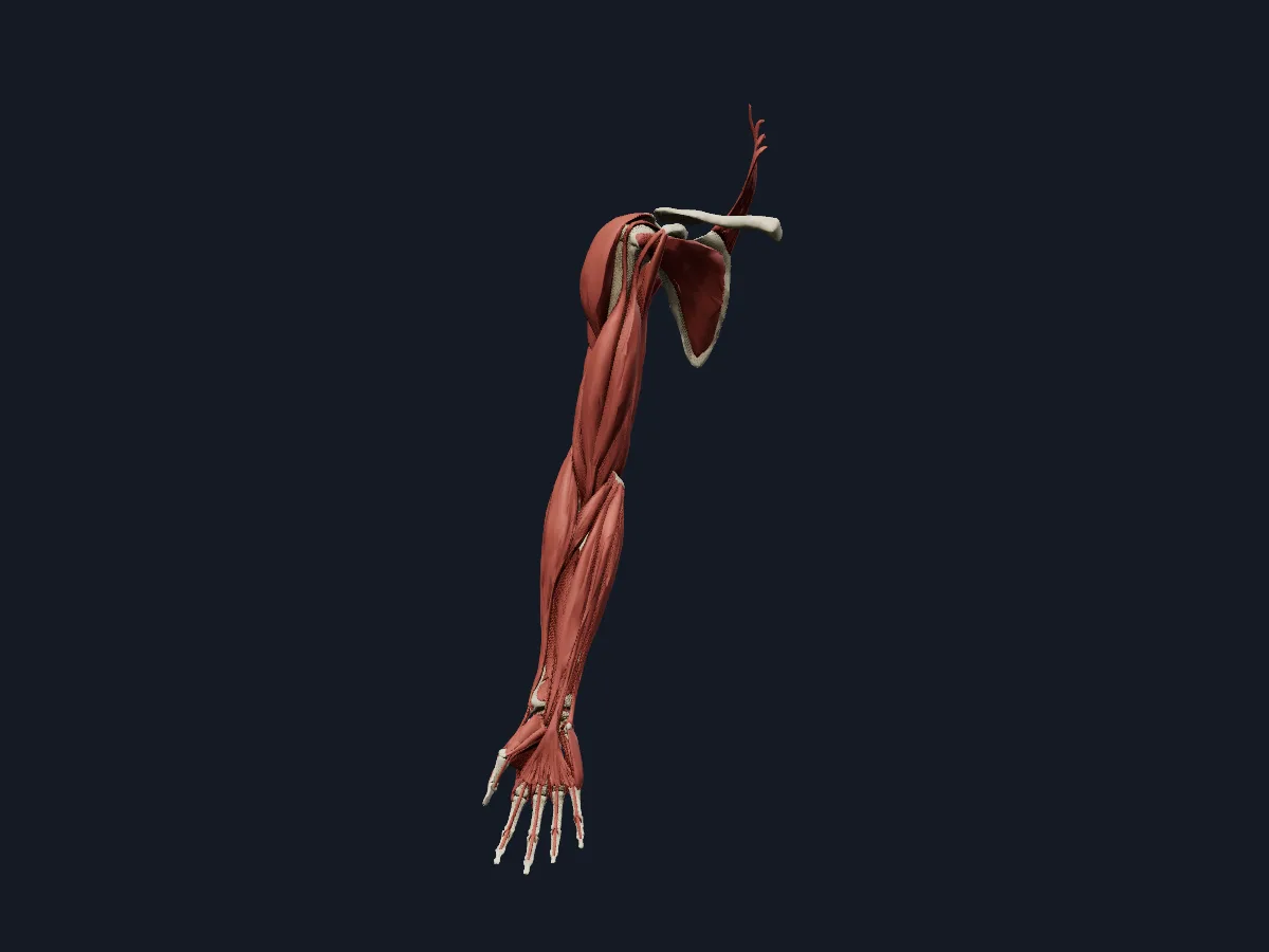 Image: Cogni Anatomy · CC BY 4.0 · Sources: BodyParts3D
- 3 muscles in the anterior compartment of the arm: biceps brachii, brachialis and coracobrachialis
- Triceps brachii has 3 heads: long, lateral and medial
- 8 muscles in the anterior (flexor) compartment of the forearm, arranged in three layers
- 12 muscles in the posterior (extensor) compartment of the forearm
- Biceps brachii is the strongest supinator of the forearm as well as an elbow flexor
Muscle groups of the upper limb
Biceps brachii and brachialis flex the elbow; coracobrachialis flexes and adducts the arm at the shoulder. All three are supplied by the musculocutaneous nerve. The posterior compartment holds triceps brachii, which extends the elbow and is supplied by the radial nerve. The deltoid caps the shoulder and abducts the arm.
In the forearm, most flexors arise from the medial epicondyle of the humerus and most extensors from the lateral epicondyle. Their long tendons cross the wrist into the hand, where the thenar and hypothenar muscles, the lumbricals and the interossei produce the fine movements of the thumb and fingers.
What the 3D model shows
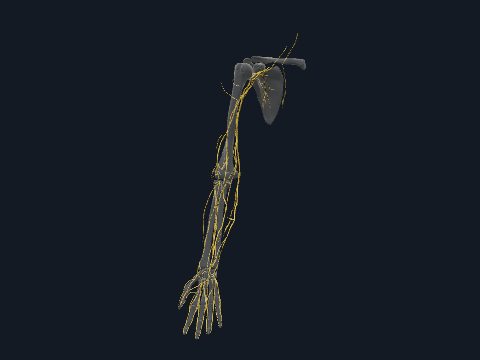
The model shows the muscles of the right upper limb on its skeleton: the acromial and spinal parts of the deltoid, subscapularis and levator scapulae; the two heads of biceps, the three heads of triceps, brachialis, coracobrachialis and anconeus; the forearm flexors and extensors, including pronator teres, supinator and pronator quadratus; and the thenar, hypothenar, lumbrical and interosseous muscles of the hand. A second view adds the arteries, veins and nerves of the limb, the interosseous membrane of the forearm and the flexor retinaculum.
How to study the arm muscles
Begin in the anterior view and find biceps brachii, then hide it to reveal brachialis underneath. Switch to the posterior view for the three heads of triceps. In the forearm, name the superficial flexors from lateral to medial: pronator teres, flexor carpi radialis, palmaris longus, flexor carpi ulnaris. Then use the muscle layers to uncover flexor digitorum superficialis and the deep layer below it. For each muscle, say its origin, insertion and action before you click it, and check the answer in the model.
Views in the 3D model
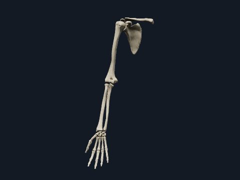
The muscles of the right upper limb on its bones, from the shoulder to the fingers.
Open this view in 3DImage: Cogni Anatomy · CC BY 4.0 · Sources: BodyParts3D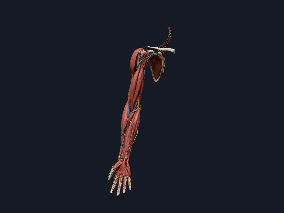
Every layer of the right upper limb together: bones, muscles, arteries, veins, nerves and connective tissue.
Open this view in 3DImage: Cogni Anatomy · CC BY-SA 4.0 · Sources: BodyParts3D, Z-Anatomy
What is in this model
185 3D parts. Names follow the Foundational Model of Anatomy (FMA).
- Muscles 47
- Arteries 42
- Nerves 35
- Bones 32
- Veins 27
- Connective tissue 2
- (Communicating branch of median nerve with ulnar nerve)
- Abductor digiti minimi of right hand
- Abductor pollicis brevis
- Abductor pollicis longus
- Acromial part of right deltoid
- Anconeus
- Anterior branch of medial antebrachial cutaneous nerve
- Anterior circumflex humeral artery
- Anterior circumflex humeral vein
- Anterior interosseous artery
- Anterior interosseous nerve of forearm
- Anterior ulnar recurrent artery
- Arteria princeps pollicis
- Arteria radialis indicis
- Basilic vein
- Brachial artery
- Brachialis
- Brachioradialis
- Capitate
- Cephalic vein
- Circumflex scapular artery
- Circumflex scapular vein
- Clavicle
- Common interosseous artery
- Common palmar digital branches of median nerve
- Common palmar digital branches of ulnar nerve
- Common palmar digital vein
- Coracobrachialis
- Deep brachial artery
- Deep branch of radial nerve
- Deep branch of ulnar nerve
- Deep palmar arch
- Deep palmar venous arch
- Distal phalanx of right index finger
- Distal phalanx of right little finger
- Distal phalanx of right middle finger
- Distal phalanx of right ring finger
- Distal phalanx of right thumb
- Dorsal branch of ulnar nerve
- Dorsal carpal branch of right radial artery
- and 133 more
Questions
What are the main muscles of the arm?
The upper arm has biceps brachii, brachialis and coracobrachialis at the front and triceps brachii at the back, with the deltoid over the shoulder. The forearm has about twenty muscles, divided into anterior flexors and posterior extensors.
Which muscle bends the elbow?
Brachialis is the main flexor of the elbow in every forearm position. Biceps brachii flexes most strongly when the forearm is supinated, and brachioradialis helps when the forearm is midway between pronation and supination.
Is this 3D arm muscle model free?
Yes. The arm muscle model and the rest of the atlas are free to use in the browser, without an account.