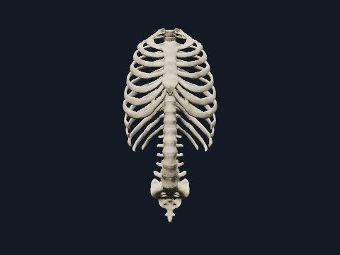
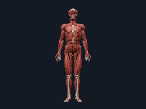
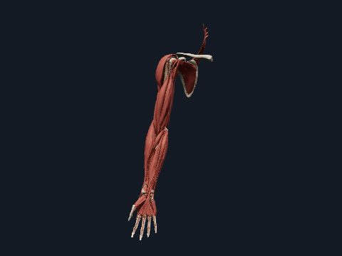
Trunk muscles in 3D
The trunk muscles move and stabilise the spine, move the ribs during breathing, anchor the shoulder girdle and close the floor of the pelvis. They include the pectoral and superficial back muscles, the intercostal muscles and diaphragm, the deep back muscles along the spine and the pelvic floor. In this free 3D model you can peel the muscles layer by layer on the skeleton and isolate any single muscle.
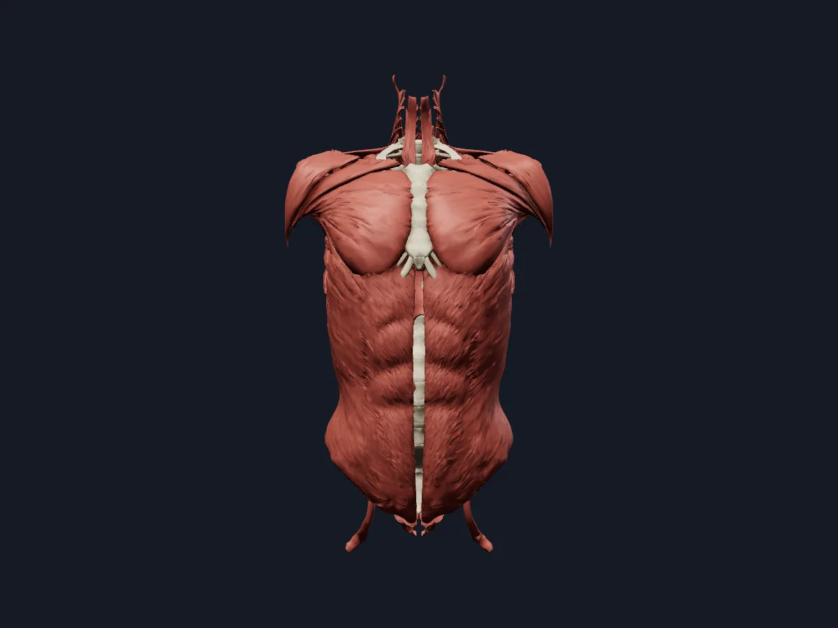 Image: Cogni Anatomy · CC BY 4.0 · Sources: BodyParts3D
- Erector spinae has 3 columns, from lateral to medial: iliocostalis, longissimus and spinalis
- 3 layers of intercostal muscles: external, internal and innermost
- The diaphragm is the main muscle of inspiration and is supplied by the phrenic nerve (C3–C5)
- Levator ani has 3 parts: puborectalis, pubococcygeus and iliococcygeus
- Pectoralis major has clavicular, sternocostal and abdominal parts
Groups of trunk muscles
On the back, the superficial layer (trapezius, rhomboids) connects the spine to the shoulder girdle. The intermediate layer, serratus posterior superior and inferior, attaches to the ribs. The deep intrinsic muscles lie in the groove beside the spinous processes: erector spinae, and below it the semispinalis, rotatores, interspinales and intertransversarii.
On the front, pectoralis major and minor, subclavius and serratus anterior cover the thoracic wall. The intercostal muscles, transversus thoracis and levatores costarum fill and move the rib cage, and the dome-shaped diaphragm separates chest from abdomen. Psoas major runs along the lumbar spine, and the pelvic floor closes the outlet of the pelvis.
What the 3D model shows
The muscles and bones view shows over a hundred muscle parts on the trunk skeleton: pectoralis major in its three parts, pectoralis minor, serratus anterior, the trapezius, rhomboids and erector spinae columns, semispinalis, splenius cervicis, supraspinatus, infraspinatus, teres major and minor, the intercostals, diaphragm, external oblique, psoas major and the levator ani, coccygeus and external anal sphincter. The all-systems view adds the vessels, nerves and organs of the trunk, more than 1,100 parts in total.
How to study the trunk muscles
Start in the posterior view and use the muscle layers control to remove one layer at a time: trapezius and rhomboids first, then the serratus posterior muscles, then the three columns of erector spinae, and finally the short muscles between the vertebrae. Switch to the anterior view, hide pectoralis major to reveal pectoralis minor and serratus anterior, and use the inferior view for the pelvic floor. For each muscle you isolate, say its attachments and main action aloud, then close the model and repeat the list from memory.
Views in the 3D model
Trunk muscles on the skeleton, from the pectoral and back muscles to the diaphragm, psoas and pelvic floor.
Open this view in 3DImage: Cogni Anatomy · CC BY 4.0 · Sources: BodyParts3D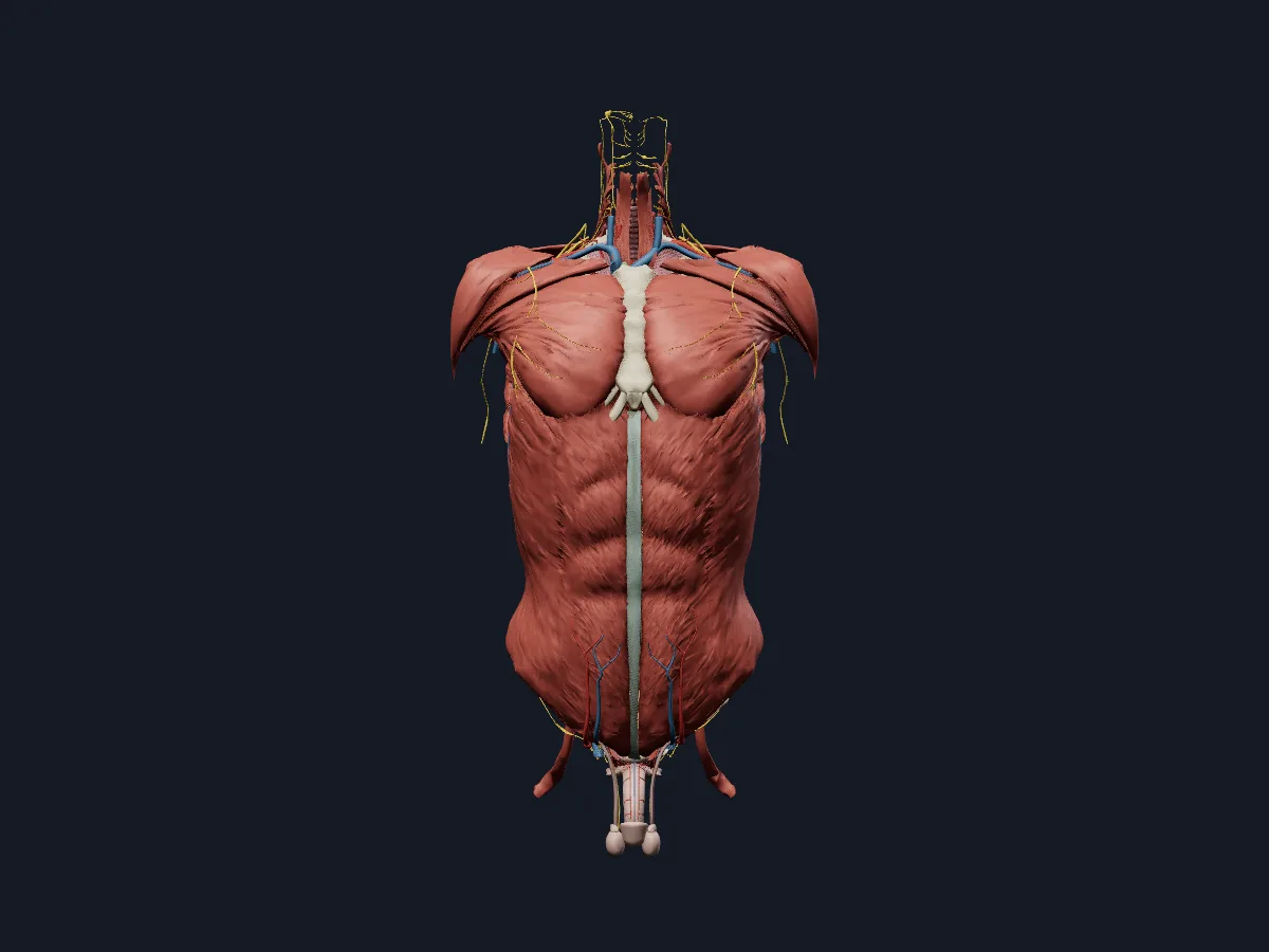
Every system of the trunk at once: bones, muscles, vessels, nerves and the thoracic, abdominal and pelvic organs.
Open this view in 3DImage: Cogni Anatomy · CC BY-SA 4.0 · Sources: BodyParts3D, HRA, Z-Anatomy
What is in this model
1144 3D parts. Names follow the Foundational Model of Anatomy (FMA).
- Arteries 339
- Veins 279
- Muscles 107
- Respiratory 106
- Digestive 101
- Nerves 89
- Bones 77
- Heart 18
- Reproductive 12
- Urinary 6
- Connective tissue 5
- Lymphatic 3
- Endocrine 2
- Abdominal aorta
- Abdominal part of left pectoralis major
- Abdominal part of right pectoralis major
- Accessory hemiazygos vein
- Accessory nerve (XI)
- Accessory subsuperior branch of right posterior basal segmental artery
- Acromial branch of left thoraco-acromial artery
- Acromial branch of right thoraco-acromial artery
- Adrenal gland
- Anterior basal segmental artery
- Anterior basal segmental bronchial tree
- Anterior basal segmental vein
- Anterior branch of lateral segmental artery
- Anterior branch of right anterior segmental artery
- Anterior cardiac vein
- Anterior cecal artery
- Anterior cusp of aortic valve
- Anterior cusp of pulmonary valve
- Anterior division of inferior trunk of brachial plexus
- Anterior division of left renal artery
- Anterior division of middle trunk of brachial plexus
- Anterior division of right renal artery
- Anterior division of superior trunk of brachial plexus
- Anterior inferior pancreaticoduodenal artery
- Anterior inferior segmental branch of right portal vein
- Anterior inferior segmental hepatic artery
- Anterior inferior segmental tributary of middle hepatic vein
- Anterior inferior tributary of right hepatic biliary tree
- Anterior interventricular vein
- Anterior leaflet of mitral valve
- Anterior leaflet of tricuspid valve
- Anterior papillary muscle of right ventricle
- Anterior part of lateral segmental vein
- Anterior part of right apical segmental artery
- Anterior part of right apical segmental vein
- Anterior root of posterior femoral cutaneous nerve
- Anterior root of spinal nerve
- Anterior segmental artery
- Anterior segmental bronchial tree
- Anterior segmental vein
- and 498 more
Questions
What are the deep back muscles?
The intrinsic back muscles that lie close to the spine and are supplied by the posterior rami of spinal nerves: splenius, erector spinae (iliocostalis, longissimus, spinalis) and the transversospinal group (semispinalis, multifidus, rotatores), plus the interspinales and intertransversarii.
Which muscles are used for breathing?
The diaphragm does most of the work of quiet inspiration, helped by the external intercostal muscles that raise the ribs. Internal and innermost intercostals, abdominal wall muscles and several neck and chest muscles assist in forced breathing.
What muscles form the pelvic floor?
The pelvic diaphragm is formed by levator ani, with its puborectalis, pubococcygeus and iliococcygeus parts, and by coccygeus. Together they support the pelvic organs.