The circulatory system in 3D
The circulatory system is the heart and the network of blood vessels that carry blood around the body: arteries take it away from the heart, capillaries exchange oxygen and nutrients with the tissues, and veins bring it back. It works as two circuits, the pulmonary circuit through the lungs and the systemic circuit through the rest of the body. In this free 3D model you can follow the arteries and veins of the whole body, from the aorta to the hands and feet.
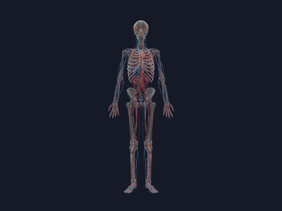 Image: Cogni Anatomy · CC BY-SA 4.0 · Sources: BodyParts3D, Z-Anatomy
- An adult has about 5 litres of blood
- At rest the heart pumps about 5 litres of blood per minute
- The aorta is the largest artery; the superior and inferior venae cavae are the largest veins
- 2 circuits: pulmonary (right heart to lungs) and systemic (left heart to the body)
- The hepatic portal vein carries blood from the gut and spleen to the liver
How the blood vessels are organised
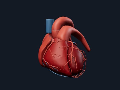
The aorta leaves the left ventricle, arches over the heart and descends through the thorax and abdomen. Its branches include the coronary, brachiocephalic, common carotid and subclavian arteries, the celiac trunk, the mesenteric and renal arteries and finally the common iliac arteries. Blood returns through the superior and inferior venae cavae to the right atrium.
Arteries have thick muscular walls that withstand high pressure; veins have thinner walls, and limb veins contain valves. Blood from the stomach, intestines and spleen does not go straight back to the heart: it first passes through the liver in the hepatic portal vein.
What the 3D model shows
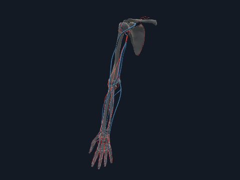
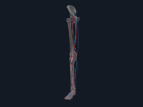
The model shows the arteries and veins of the whole body with the skeleton as see-through context: the aorta and its branches, the coronary arteries and cardiac veins, the pulmonary trunk and pulmonary veins, the cerebral arteries that form the arterial circle at the base of the brain, the dural venous sinuses, the jugular, subclavian and axillary vessels, the azygos system, the vessels of the stomach, liver, intestines and kidneys, the hepatic portal vein, and the vessels of both arms and legs down to the digits. Separate views show arteries only or veins only.
How to study the circulatory system
Open the arteries view and follow the aorta from the heart: name the three branches of the arch, then the unpaired branches of the abdominal aorta from top to bottom, then the paired renal and gonadal arteries. Continue into one arm and one leg. Switch to the veins view and trace blood back from the head through the internal jugular vein, and from the gut through the portal vein to the liver. Recall each route aloud the next day before opening the model again.
Views in the 3D model
Arteries and veins of the whole body together, over a see-through skeleton.
Open this view in 3DImage: Cogni Anatomy · CC BY-SA 4.0 · Sources: BodyParts3D, Z-Anatomy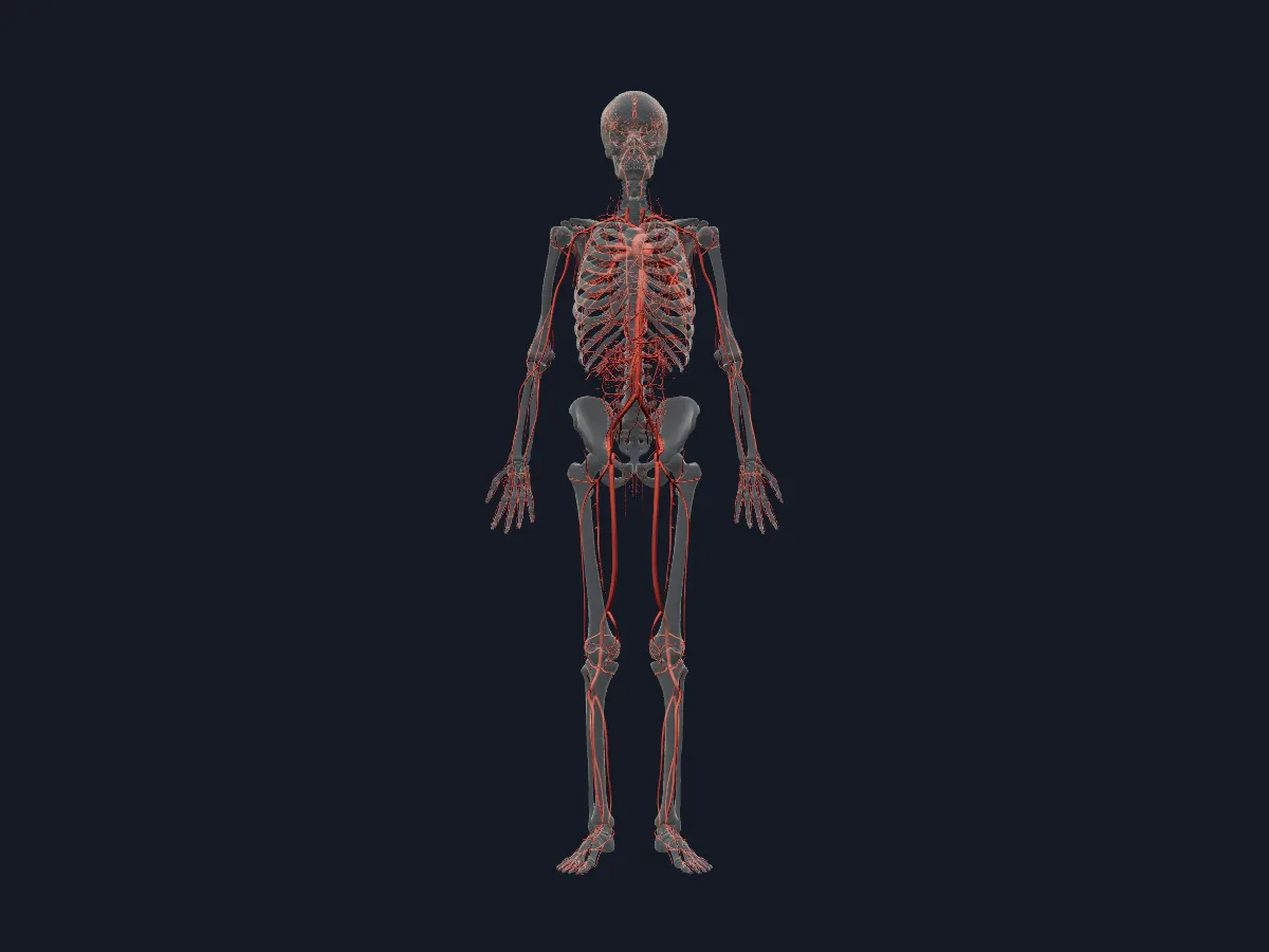
The arterial tree of the whole body, from the aorta and the arteries of the brain to the hands and feet.
Open this view in 3DImage: Cogni Anatomy · CC BY 4.0 · Sources: BodyParts3D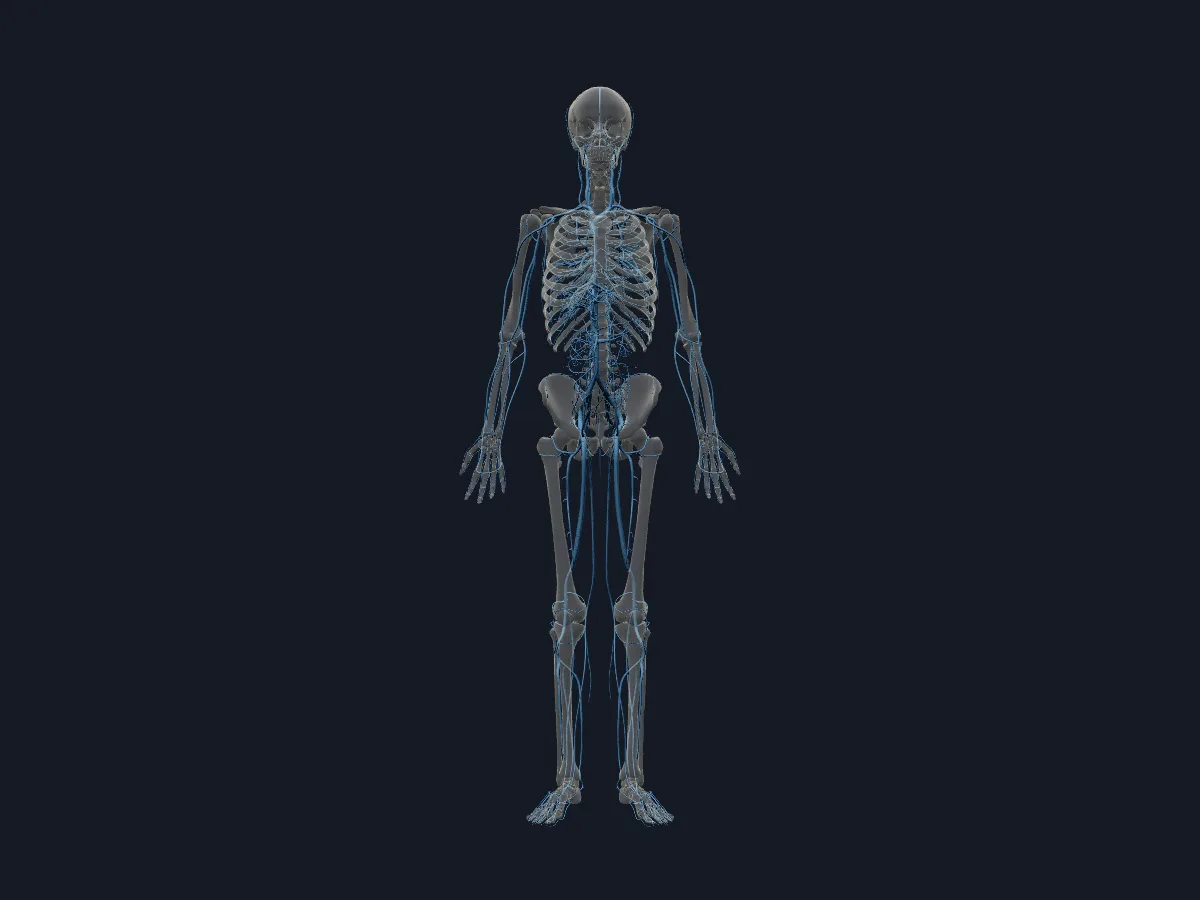
The veins of the whole body, including the venae cavae, the dural sinuses and the hepatic portal system.
Open this view in 3DImage: Cogni Anatomy · CC BY-SA 4.0 · Sources: BodyParts3D, Z-Anatomy
What is in this model
1091 3D parts. Names follow the Foundational Model of Anatomy (FMA).
- Arteries 639
- Veins 452
- Abdominal aorta
- Accessory hemiazygos vein
- Accessory subsuperior branch of right posterior basal segmental artery
- Acromial branch of left thoraco-acromial artery
- Acromial branch of right thoraco-acromial artery
- Angular vein
- Anterior basal segmental artery
- Anterior basal segmental vein
- Anterior branch of lateral segmental artery
- Anterior branch of right anterior segmental artery
- Anterior cardiac vein
- Anterior cecal artery
- Anterior cerebral artery
- Anterior choroidal artery
- Anterior circumflex humeral artery
- Anterior circumflex humeral vein
- Anterior communicating artery
- Anterior division of left renal artery
- Anterior division of retromandibular vein
- Anterior division of right renal artery
- Anterior inferior cerebellar artery
- Anterior inferior pancreaticoduodenal artery
- Anterior inferior segmental branch of right portal vein
- Anterior inferior segmental hepatic artery
- Anterior inferior segmental tributary of middle hepatic vein
- Anterior intercavernous sinus
- Anterior interosseous artery
- Anterior interventricular vein
- Anterior parietal artery
- Anterior part of lateral segmental vein
- Anterior part of right apical segmental artery
- Anterior part of right apical segmental vein
- Anterior segmental artery
- Anterior segmental vein
- Anterior spinal artery
- Anterior superior pancreaticoduodenal artery
- Anterior superior segmental branch of right portal vein
- Anterior superior segmental hepatic artery
- Anterior superior segmental tributary of right hepatic vein
- Anterior temporal artery
- and 459 more
Questions
What is the difference between arteries and veins?
Arteries carry blood away from the heart and have thick, muscular walls; veins carry blood back to the heart, have thinner walls and, in the limbs, valves. In the pulmonary circuit the pulmonary arteries carry deoxygenated blood and the pulmonary veins oxygenated blood, the reverse of the systemic circuit.
What is the largest artery in the body?
The aorta. It begins at the left ventricle, forms the aortic arch and descends through the thorax and abdomen before dividing into the common iliac arteries at about the level of the fourth lumbar vertebra.
How much blood is in the human body?
About 5 litres in an average adult, roughly 7% of body weight.