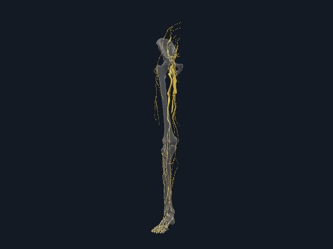
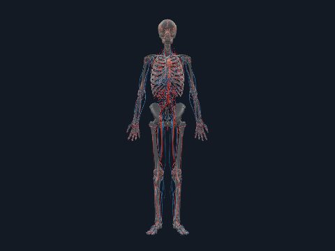
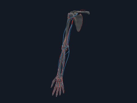
Blood vessels of the leg in 3D
Blood reaches the leg (lower limb) through the external iliac artery, which becomes the femoral artery at the groin and the popliteal artery behind the knee before dividing into the anterior and posterior tibial arteries. It returns through deep veins that run with these arteries and through the great and small saphenous veins under the skin. In this free 3D model you can trace the arteries and veins of the right lower limb over a see-through skeleton.
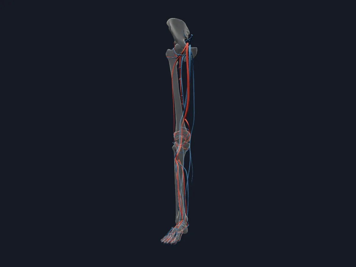 Image: Cogni Anatomy · CC BY 4.0 · Sources: BodyParts3D
- The external iliac artery becomes the femoral artery as it passes under the inguinal ligament
- The femoral artery becomes the popliteal artery at the adductor hiatus
- The anterior tibial artery continues on the foot as the dorsalis pedis artery
- The great saphenous vein is the longest vein in the body
- The small saphenous vein passes behind the lateral malleolus and drains into the popliteal vein
Arteries and veins of the lower limb
In the thigh the femoral artery runs through the femoral triangle and the adductor canal, and its circumflex and perforating branches supply the thigh muscles. Genicular arteries from the popliteal artery join in an anastomosis around the knee. The anterior tibial artery passes forward above the interosseous membrane; the posterior tibial artery runs behind the medial malleolus and divides into the medial and lateral plantar arteries, and the lateral one forms the plantar arch.
The great saphenous vein starts at the medial end of the dorsal venous arch, passes in front of the medial malleolus and ends in the femoral vein. Perforating veins connect the superficial and deep systems.
What the 3D model shows
The model shows the arteries and veins of the right lower limb with the skeleton as see-through context. Arteries run from the external and internal iliac arteries through the femoral, lateral circumflex femoral, perforating and popliteal arteries with their genicular branches to the anterior and posterior tibial arteries, the dorsalis pedis, arcuate and plantar arteries and the plantar arch. Veins include the great and small saphenous veins, the femoral, deep femoral, popliteal, tibial and fibular veins, the perforating veins and the dorsal and plantar venous arches. Separate views show the arteries and the veins alone.
How to study the vessels of the leg
Open the arteries view and follow one path from the external iliac artery to the toes, naming the landmark where each vessel changes name: the inguinal ligament, the adductor hiatus, the lower border of popliteus, the ankle. Rotate to the posterior view for the popliteal artery and its genicular branches. In the veins view, trace the great saphenous vein up the medial side and the small saphenous vein up the back of the calf. Then show both systems together and pair each deep vein with its artery.
Views in the 3D model
Arteries and veins of the right lower limb together, over a see-through skeleton.
Open this view in 3DImage: Cogni Anatomy · CC BY 4.0 · Sources: BodyParts3D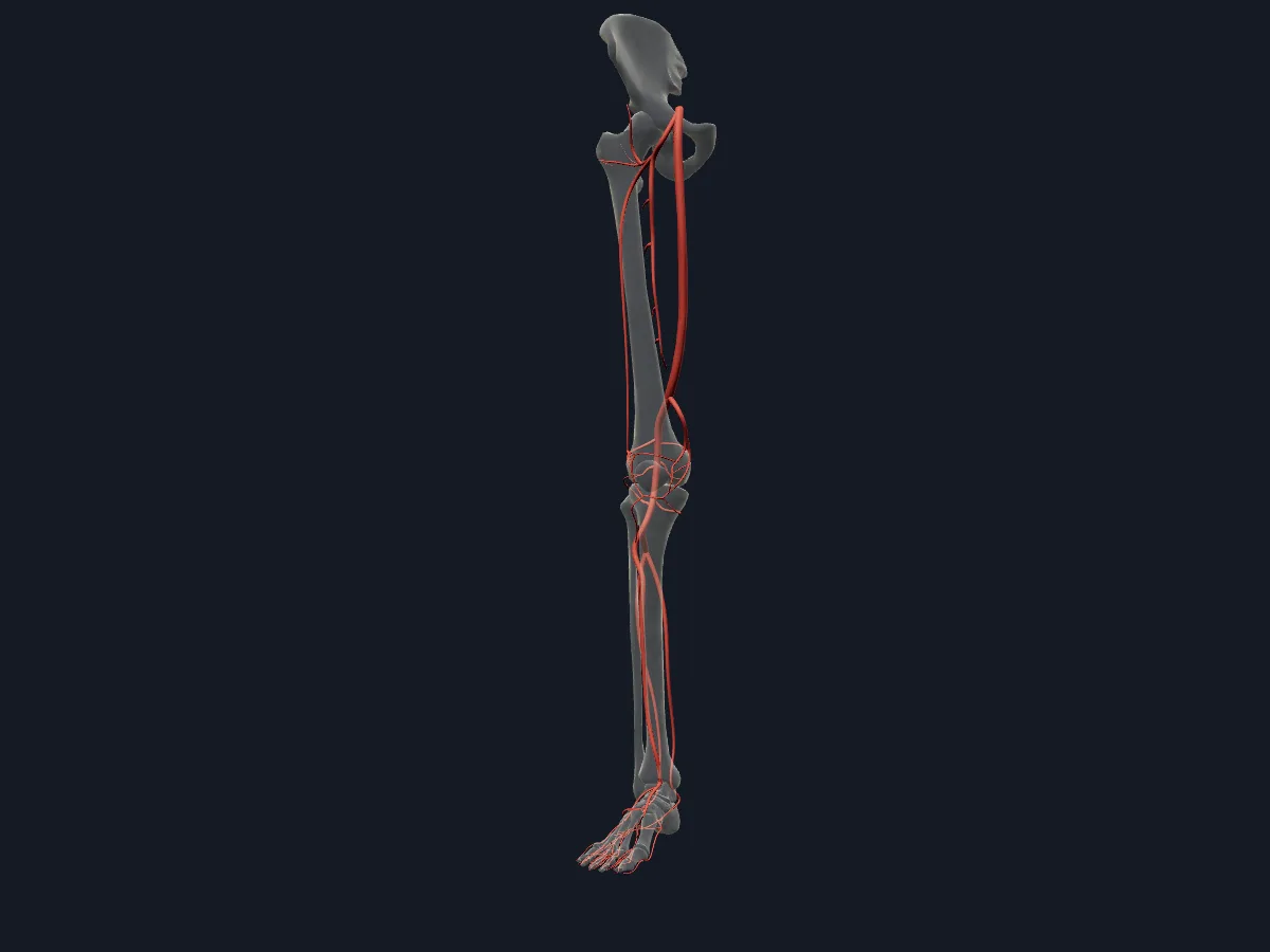
The arteries of the right lower limb from the iliac arteries to the plantar arch and toes.
Open this view in 3DImage: Cogni Anatomy · CC BY 4.0 · Sources: BodyParts3D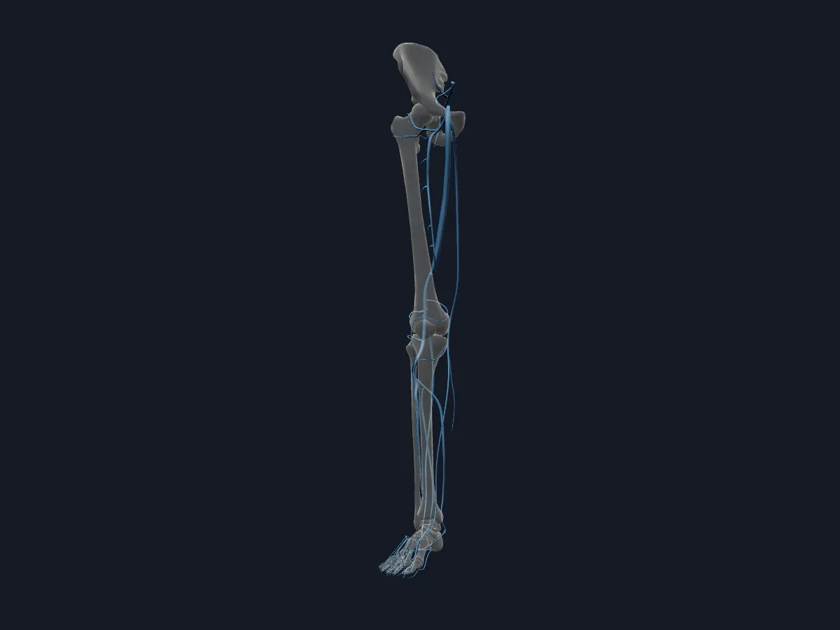
The deep and superficial veins of the right lower limb, including the great and small saphenous veins.
Open this view in 3DImage: Cogni Anatomy · CC BY 4.0 · Sources: BodyParts3D
What is in this model
62 3D parts. Names follow the Foundational Model of Anatomy (FMA).
- Arteries 31
- Veins 31
- Anterior tibial artery
- Anterior tibial recurrent artery
- Anterior tibial vein
- Arcuate artery
- Deep femoral vein
- Deep plantar artery
- Descending branch of right lateral circumflex femoral artery
- Descending genicular artery
- Distal perforating artery
- Dorsal digital artery of foot
- Dorsal metacarpal vein
- Dorsal venous arch of right foot
- Dorsalis pedis artery
- Femoral artery
- Femoral vein
- Fibular vein
- Genicular vein
- Great saphenous vein
- Inferior gluteal vein
- Inferior lateral genicular artery
- Inferior medial genicular artery
- Lateral circumflex femoral artery
- Lateral circumflex femoral vein
- Lateral plantar artery
- Lateral superior genicular artery
- Lateral tarsal artery
- Medial circumflex femoral vein
- Medial plantar artery
- Medial superior genicular artery
- Middle genicular artery
- Obturator vein
- Plantar arch
- Plantar metatarsal artery
- Plantar metatarsal vein
- Plantar venous arch of right foot
- Popliteal artery
- Popliteal vein
- Posterior tibial artery
- Posterior tibial vein
- Set of calcaneal branches of posterior tibial artery
- and 11 more
Questions
What is the main artery of the leg?
The femoral artery, the continuation of the external iliac artery. Behind the knee it becomes the popliteal artery, which divides into the anterior and posterior tibial arteries.
What is the longest vein in the body?
The great saphenous vein. It runs from the dorsal venous arch of the foot up the medial side of the leg and thigh and joins the femoral vein just below the groin.
Where can the pulses of the leg be felt?
The femoral pulse below the inguinal ligament, the popliteal pulse deep in the popliteal fossa, the posterior tibial pulse behind the medial malleolus, and the dorsalis pedis pulse on the top of the foot, lateral to the tendon of extensor hallucis longus.