Heart anatomy in 3D
The heart is a four-chambered muscular pump in the middle of the chest that drives blood through the lungs and the rest of the body. In this free 3D model you can turn the heart around, open it to see the atria, ventricles and valves, and follow the coronary arteries on its surface.
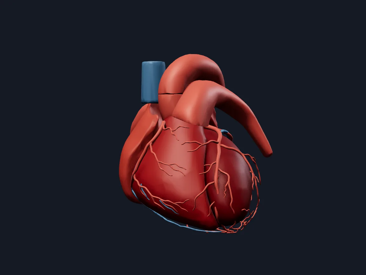 Image: Cogni Anatomy · CC BY 4.0 · Sources: BodyParts3D, HRA
- 4 chambers: right and left atria, right and left ventricles
- 4 valves: tricuspid, pulmonary, mitral and aortic
- About the size of a clenched fist, roughly 250–350 g in adults
- Lies in the middle mediastinum, about two-thirds of it left of the midline
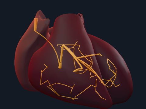
- The left and right coronary arteries arise from the aortic root and supply the heart wall
Structure of the heart
The right atrium receives deoxygenated blood from the superior and inferior venae cavae and the coronary sinus and passes it through the tricuspid valve to the right ventricle, which pumps it into the pulmonary trunk. Oxygenated blood returns through the pulmonary veins to the left atrium, crosses the mitral valve into the left ventricle and leaves through the aortic valve into the aorta.
The interatrial and interventricular septa separate the two sides. The wall has three layers, epicardium, myocardium and endocardium, and the left ventricular wall is about three times thicker than the right because it pumps against systemic pressure. The whole heart sits in the pericardial sac.
What the 3D model shows
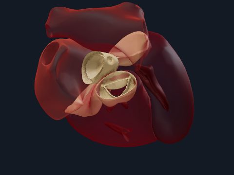
The external view shows the heart with the aorta, pulmonary trunk, pulmonary arteries and superior vena cava, and the coronary arteries and cardiac veins on its surface. The chambers view colours each atrium and ventricle separately, so you can see how the right ventricle wraps around the front of the left. The detailed teaching model opens the heart to show the valves, chordae tendineae and papillary muscles.
How to study the heart
Start from the anterior view: the right ventricle forms most of the front surface and the left ventricle the apex. Turn to the posterior view to find the left atrium and the coronary sinus. Then open the heart and trace one drop of blood from the venae cavae to the aorta, naming every chamber and valve on the way. Recalling the path from memory the next day fixes it far better than rereading.
Views in the 3D model
External view with the great vessels and the coronary arteries and veins on the surface.
Open this view in 3DImage: Cogni Anatomy · CC BY 4.0 · Sources: BodyParts3D, HRA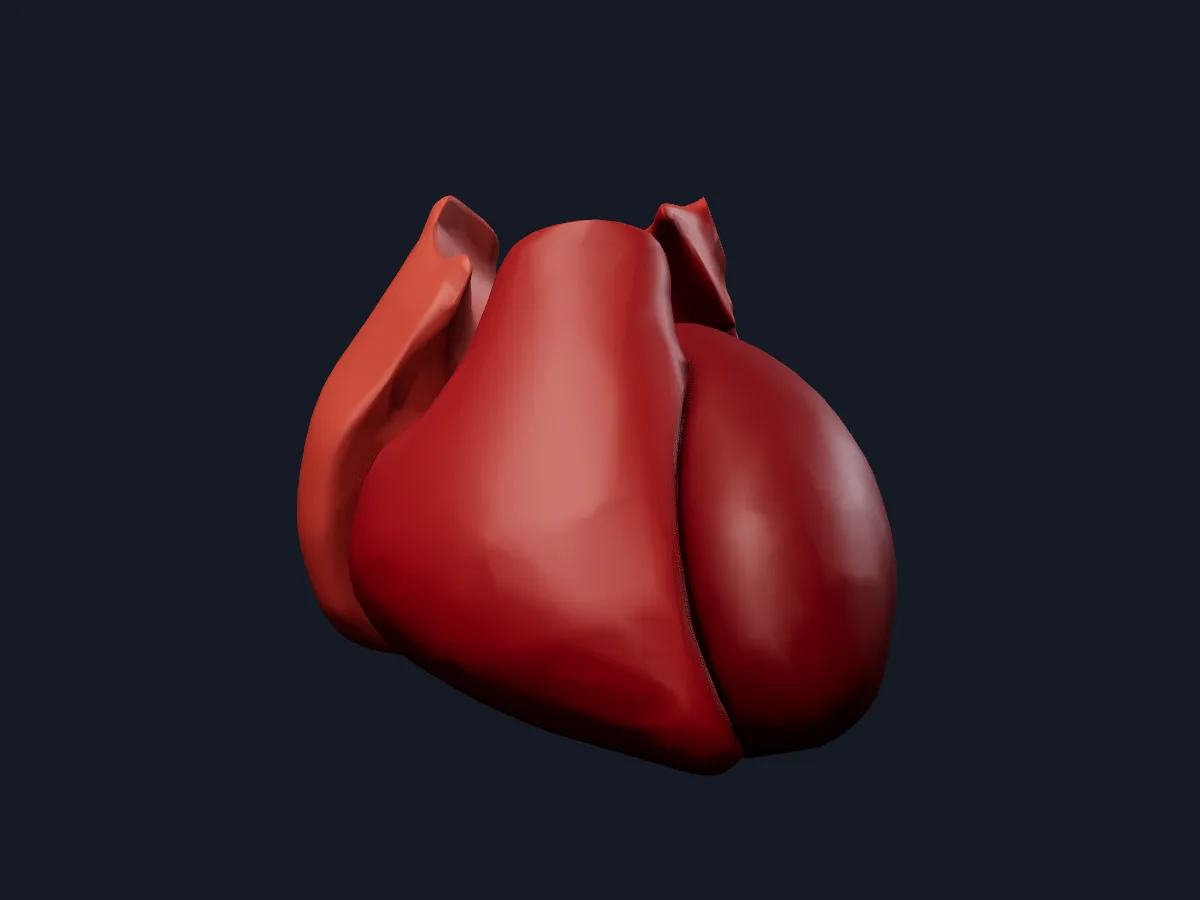
Atria and ventricles in separate colours, showing how the four chambers fit together.
Open this view in 3DImage: Cogni Anatomy · CC BY 4.0 · Sources: HRA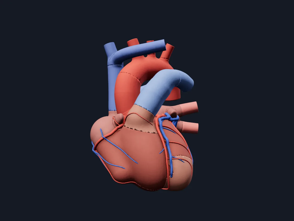
A detailed teaching model of the heart walls, valves, chordae and papillary muscles.
Open this view in 3DImage: Cogni Anatomy · MIT · Sources: Layered Human
What is in this model
202 3D parts. Names follow the Foundational Model of Anatomy (FMA).
- Arteries 83
- Heart 83
- Veins 36
- Anterior descending artery
- Aortic arch
- Aortic left coronary cusp
- Aortic non coronary cusp
- Aortic right coronary cusp
- Aortic valve
- Arch of aorta
- Ascending aorta
- Atrioventricular nodal artery
- Atrium anterior wall
- Atrium lateral wall
- Atrium posterior wall
- Auricle
- Brachiocephalic trunk
- Brachiocephalic vein
- Cardiac atrium
- Central fibrous body
- Circumflex artery
- Circumflex branch of left coronary artery
- Common carotid artery
- Conus artery
- Conus branch of anterior interventricular branch of left coronary artery
- Coronary artery
- Coronary sinus
- Descending aorta
- Diagonal branch of anterior descending branch of left coronary artery
- First anterior cardiac vein
- First anterior ventricular branch of right coronary artery
- First diagonal artery
- First obtuse marginal artery
- First posterior ventricular branch of right coronary artery
- First right anterior branch of anterior interventricular branch of left coronary artery
- First septal perforator
- Great cardiac vein
- Great cardiac vein tributary 1
- Great cardiac vein tributary 2
- Great cardiac vein tributary 3
- Great cardiac vein tributary 4
- Heart left ventricle
- Heart right ventricle
- and 99 more
Questions
How many chambers does the human heart have?
Four: two atria that receive blood and two ventricles that pump it out. The right side sends blood to the lungs, the left side to the rest of the body.
Where is the heart located?
In the middle mediastinum of the chest, behind the sternum and between the lungs, with its apex pointing down and to the left at about the fifth intercostal space.
Is this 3D heart model free?
Yes. The heart model and the rest of the atlas are free to use in the browser, without an account.