Female reproductive system in 3D
The female reproductive system consists of the ovaries, uterine tubes, uterus, vagina and external genitalia, with the mammary glands as accessory organs. The ovaries produce eggs and hormones, the uterine tubes carry the egg towards the uterus, and the uterus holds a developing pregnancy. In this free 3D model you can see the ovaries, tubes and uterus with the urinary organs in the pelvis, and the internal structure of the breast.
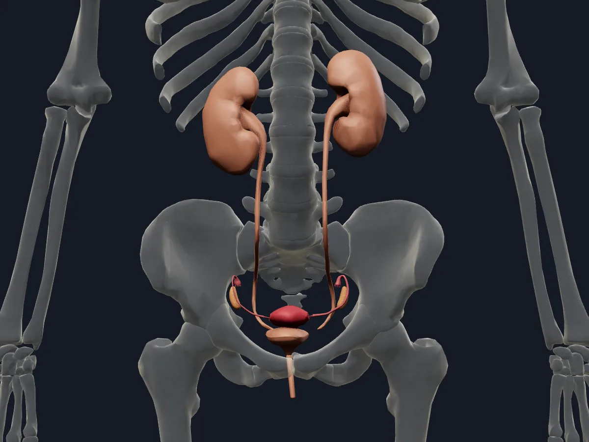 Image: Cogni Anatomy · CC BY 4.0 · Sources: BodyParts3D, HRA, Cogni
- The uterine tube is about 10 cm long, with 4 parts: infundibulum, ampulla, isthmus and uterine (intramural) part
- The uterus has a fundus, body and cervix and in adults is about 7–8 cm long
- Each ovary is almond-shaped, about 3 cm long
- The female urethra is about 4 cm long
- The mammary gland contains glandular lobes drained by lactiferous ducts that open on the nipple
Structure of the female reproductive organs
The uterus lies in the lesser pelvis between the urinary bladder in front and the rectum behind, usually tilted forward over the bladder. Its fundus is the rounded top above the openings of the uterine tubes. Each tube opens near the ovary through a funnel-shaped infundibulum fringed with fimbriae, widens into the ampulla, where fertilisation usually occurs, and narrows into the isthmus before entering the uterine wall.
The breast lies on the pectoral fascia. Its glandular lobes drain through lactiferous ducts that carry milk to the nipple, and suspensory ligaments run from the skin to the deep fascia.
What the 3D model shows
The urogenital view shows the female pelvic organs from the Human Reference Atlas: the uterus with its fundus, body and anterior and posterior walls; the uterine tubes with infundibulum, fimbriae, abdominal ostium, ampulla and isthmus; and the ovaries. The urinary organs are included: kidneys, ureters, bladder with fundus, trigone, neck and ureteral orifices, and the female urethra. The breast view shows the right breast with nipple, areola, areolar tubercles, mammary lobes, lactiferous ducts and sinuses, and suspensory ligaments. The model includes structures labelled as lactiferous sinuses, following a traditional anatomical scheme; these should not be interpreted as constant milk-storage reservoirs in the lactating breast.
How to study the female reproductive organs
Start with the urogenital view in the anterior view, then turn to the lateral view to see the uterus resting over the bladder. In the isolated view, follow the route of an egg: ovary, fimbriae, infundibulum, ampulla, isthmus, uterus. Use see-through mode on the bladder to find the trigone between the ureteral orifices. In the breast view, trace one lobe through its duct to the nipple. Then name every part from memory before checking the labels.
Views in the 3D model
Female reproductive and urinary organs in the pelvis, inside a see-through skeleton.
Open this view in 3DImage: Cogni Anatomy · CC BY 4.0 · Sources: BodyParts3D, HRA, Cogni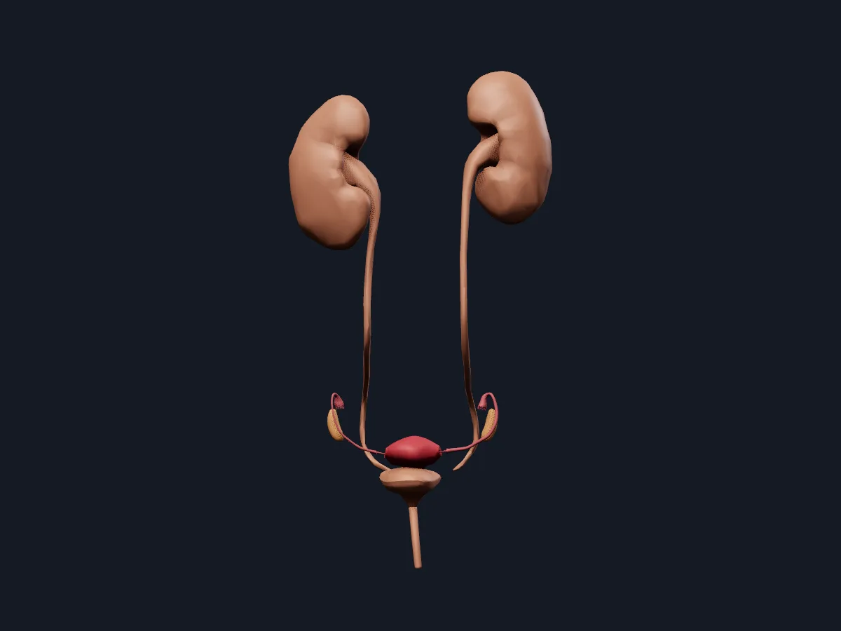
The uterus, uterine tubes, ovaries and urinary organs on their own, without surrounding structures.
Open this view in 3DImage: Cogni Anatomy · CC BY 4.0 · Sources: BodyParts3D, HRA, Cogni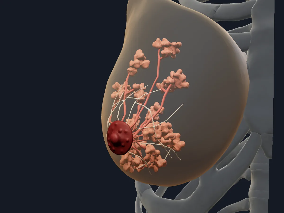
The right breast with its lobes, lactiferous ducts, nipple, areola and suspensory ligaments; labelled sinuses follow the model's traditional scheme.
Open this view in 3DImage: Cogni Anatomy · CC BY 4.0 · Sources: BodyParts3D, HRA
What is in this model
33 3D parts. Names follow the Foundational Model of Anatomy (FMA).
- Reproductive 22
- Urinary 11
- Abdominal ostium of uterine tube
- Ampulla of uterine tube
- Anterior wall of uterus
- Areola
- Areolar tubercles
- Body of uterus
- Female urethra
- Fimbria of uterine tube
- Fundus of urinary bladder base
- Fundus of urinary bladder dome
- Fundus of uterus
- Isthmus of fallopian tube
- Kidney
- Main lactiferous ducts
- Main lactiferous sinuses
- Mammary lobes
- Nipple
- Ovary
- Posterior wall of uterus
- Suspensory ligaments
- Trigone of urinary bladder
- Ureter
- Ureteral orifice
- Urinary bladder neck smooth muscle
- Uterine tube infundibulum
Questions
What are the parts of the fallopian tube?
From the ovary towards the uterus: the infundibulum with its fimbriae, the ampulla, the isthmus and the uterine part that passes through the uterine wall.
Where does fertilisation usually take place?
In the ampulla of the uterine tube, its widest and longest part. The fertilised egg then moves through the isthmus into the uterus.
What is the internal structure of the breast?
Glandular lobes separated by fat and connective tissue, lactiferous ducts that carry milk to the nipple, and suspensory ligaments that support the gland.