Kidney anatomy in 3D
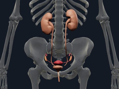
The kidneys are a pair of bean-shaped organs on the posterior abdominal wall, behind the peritoneum, that filter the blood, regulate water and salt balance and produce urine. Each kidney has a convex lateral border and a medial hilum where the renal vessels and the renal pelvis enter and leave. In this free 3D model you can see both kidneys in place, rotate them and relate them to the rest of the urinary tract.
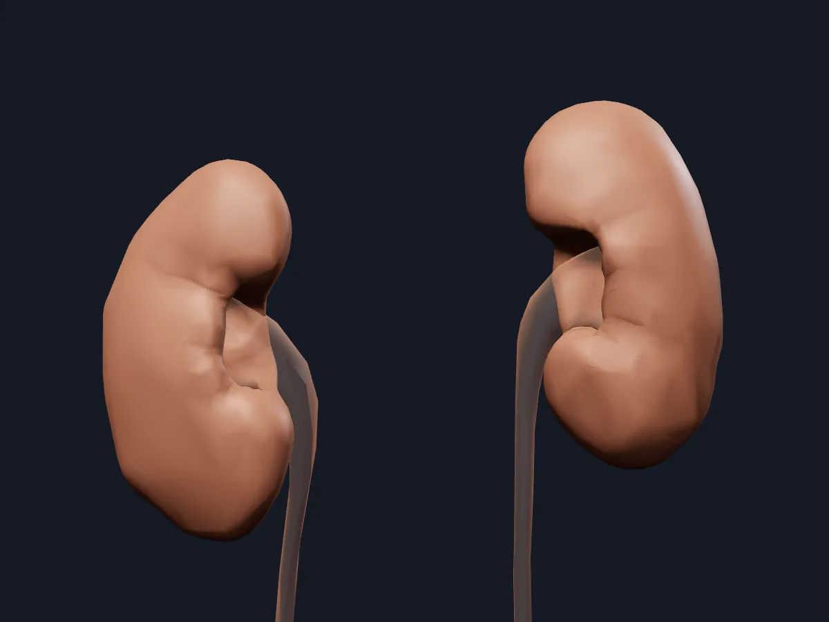 Image: Cogni Anatomy · CC BY 4.0 · Sources: BodyParts3D
- Each kidney is about 10–12 cm long, 5–6 cm wide and 3 cm thick
- The kidneys lie retroperitoneally at about T12–L3; the right kidney is usually slightly lower than the left
- About 1 million nephrons in each kidney
- The kidneys receive about 20–25% of the cardiac output at rest
- At the hilum, from front to back: renal vein, renal artery, renal pelvis
Structure of the kidney
A fibrous capsule covers the kidney, and perirenal fat and renal fascia surround it. Inside, the outer cortex contains the renal corpuscles, and the inner medulla is made of 8–18 renal pyramids separated by renal columns. Urine leaves the tip of each pyramid into a minor calyx; minor calyces join into major calyces, which form the renal pelvis, and the renal pelvis narrows into the ureter.
Behind each kidney lie the diaphragm, psoas major and quadratus lumborum, and the twelfth rib crosses its upper part. A suprarenal gland sits on each upper pole.
What the 3D model shows
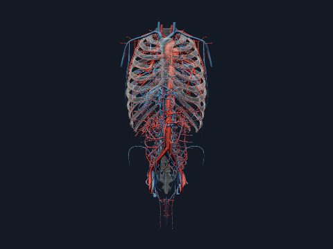
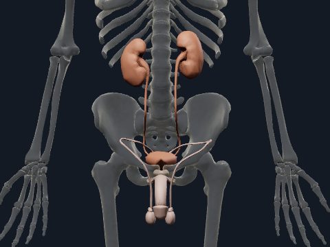
The kidney view shows the left and right kidneys as whole organs in their position on the posterior abdominal wall, with the ureters and bladder of the urinary tract faded for context. The model shows the outer form of each kidney, its poles, borders and hilum; the cortex, medulla and calyces inside are not modelled. The renal arteries and veins with their segmental branches are on the trunk blood vessels page.
How to study the kidney
Rotate the kidneys from the anterior to the posterior view and note that the hilum faces medially and slightly forward. Compare the heights of the two upper poles to see the lower position of the right kidney. Follow the faded ureter down from each hilum to the bladder. Then open the arteries view of the trunk vessels and trace the renal arteries from the aorta into the hilum. Afterwards, list the structures at the hilum and the layers inside the kidney from memory.
What is in this model
2 3D parts. Names follow the Foundational Model of Anatomy (FMA).
- Urinary 2
- Kidney
Questions
Where are the kidneys located?
On the posterior abdominal wall on either side of the vertebral column, behind the peritoneum, at about the level of T12 to L3. The upper poles are protected by the lowest ribs.
Why is the right kidney lower than the left?
The large right lobe of the liver lies above it, so the right kidney usually sits slightly lower than the left.
What does the kidney do?
It filters the blood through about a million nephrons, adjusts water, salt and acid-base balance and forms urine. It also produces hormones such as renin and erythropoietin.