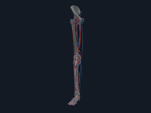
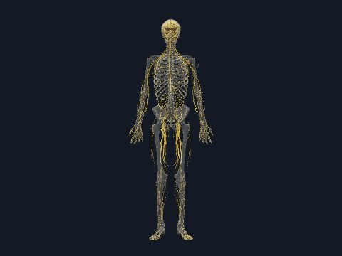
Nerves of the leg in 3D
The nerves of the leg (lower limb) come from the lumbar plexus (L1–L4) and the sacral plexus (L4–S4). The femoral and obturator nerves supply the front and inner side of the thigh, while the sciatic nerve and its tibial and common fibular branches supply the back of the thigh, the leg and the foot. In this free 3D model you can follow the nerves of the right lower limb and show them with the muscles they supply.
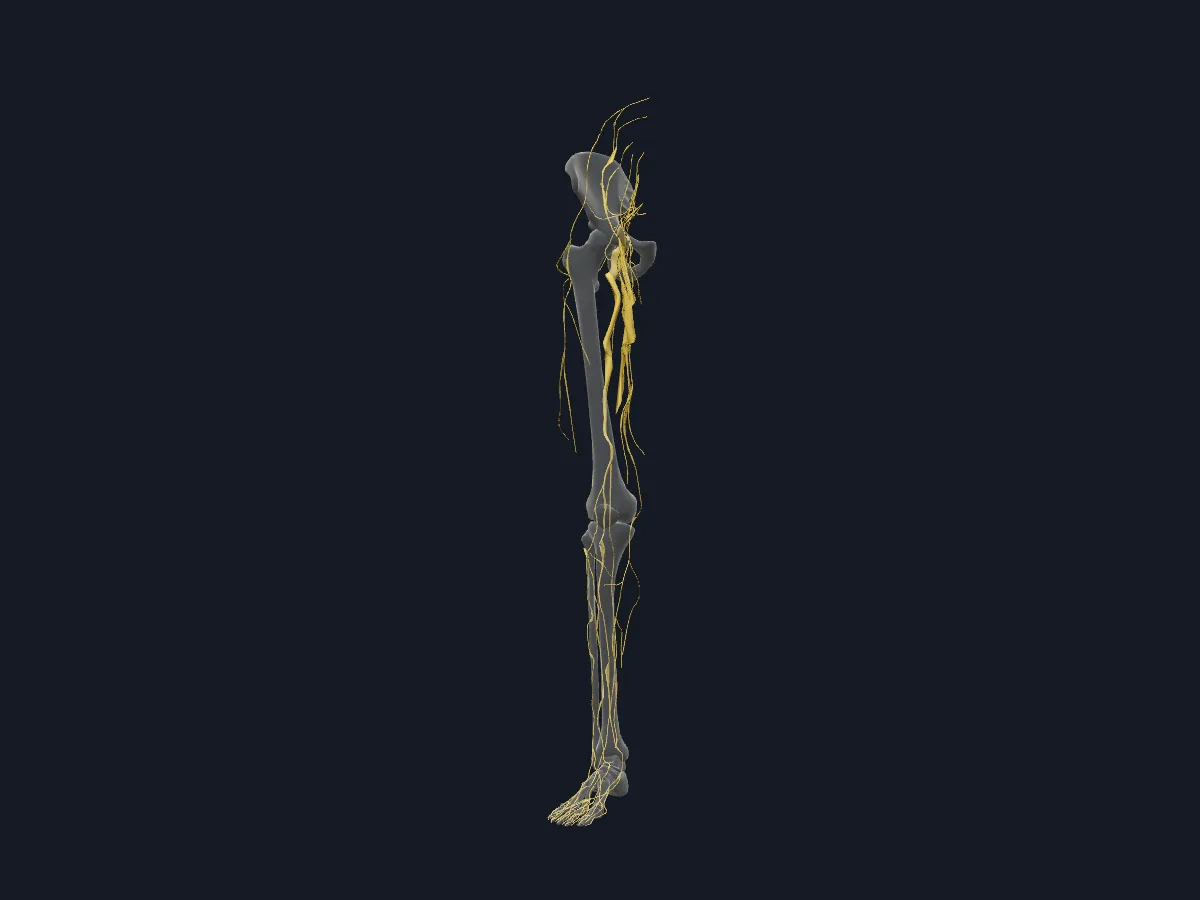 Image: Cogni Anatomy · CC BY-SA 4.0 · Sources: BodyParts3D, Z-Anatomy
- The sciatic nerve is the largest nerve in the body, formed from the L4–S3 roots
- The sciatic nerve divides into the tibial and common fibular nerves, usually above the popliteal fossa
- The femoral nerve (L2–L4) supplies the quadriceps; the obturator nerve (L2–L4) supplies most adductors
- The common fibular nerve winds around the neck of the fibula before dividing into deep and superficial branches
Course of the main nerves
The femoral nerve enters the thigh lateral to the femoral artery and splits into muscular and anterior cutaneous branches; its saphenous branch continues down the medial side of the leg. The obturator nerve leaves the pelvis through the obturator canal and divides into anterior and posterior branches around adductor brevis.
The sciatic nerve leaves the pelvis below piriformis and descends deep to the hamstrings. The tibial nerve passes through the popliteal fossa and the calf and divides behind the medial malleolus into the medial and lateral plantar nerves. The common fibular nerve gives the deep fibular nerve to the anterior compartment and the superficial fibular nerve to the lateral compartment and the skin of the dorsum of the foot.
What the 3D model shows
The model shows the nerves of the right lower limb with the skeleton as see-through context: the femoral nerve with its saphenous and anterior cutaneous branches; the anterior and posterior branches of the obturator nerve; the superior gluteal nerve and the nerves to piriformis and quadratus femoris; the sciatic, tibial and common fibular nerves; the deep and superficial fibular nerves; the sural nerve; the medial and lateral plantar nerves; and cutaneous nerves including the lateral and posterior femoral cutaneous nerves and the branches of the genitofemoral nerve. A second view adds the limb muscles.
How to study the nerves of the leg
Start in the posterior view and follow the sciatic nerve from below piriformis down the thigh to its division. Trace the tibial nerve through the calf to the sole and the common fibular nerve around the neck of the fibula. Turn to the anterior view for the femoral nerve and its saphenous branch. Then switch to the nerves-and-muscles view and work compartment by compartment: name the muscles and the nerve that supplies them, and check your answer by isolating that nerve.
Views in the 3D model
The nerves of the right lower limb and their branches, over a see-through skeleton.
Open this view in 3DImage: Cogni Anatomy · CC BY-SA 4.0 · Sources: BodyParts3D, Z-Anatomy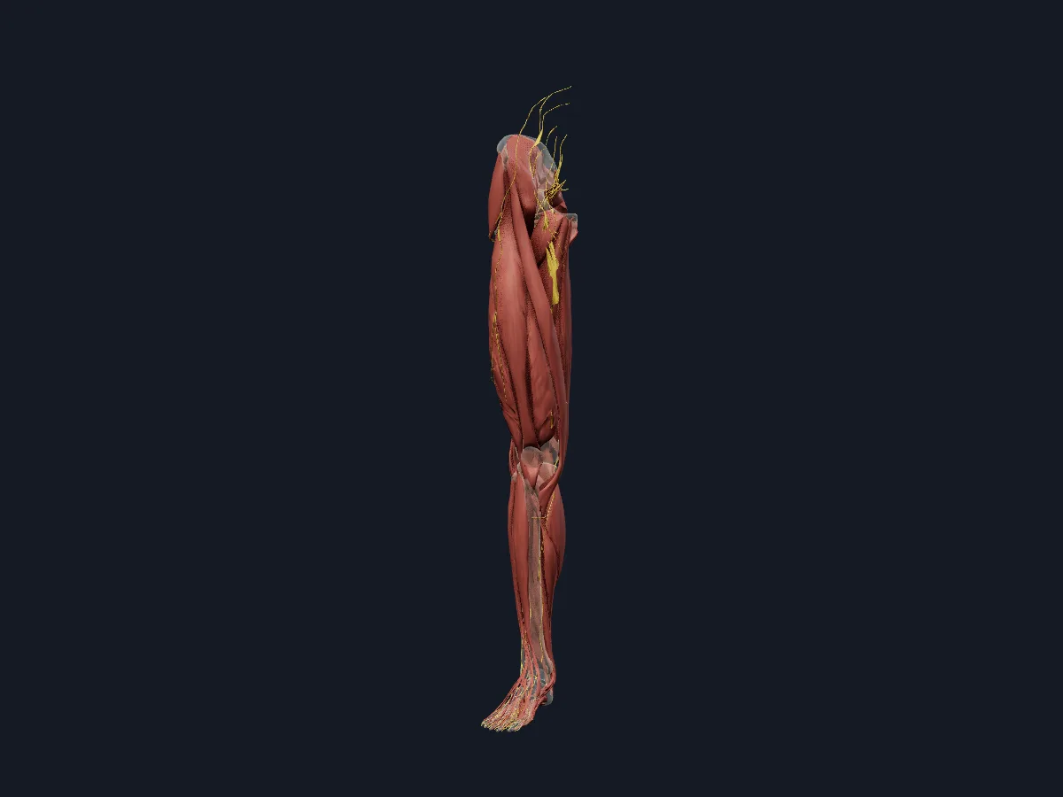
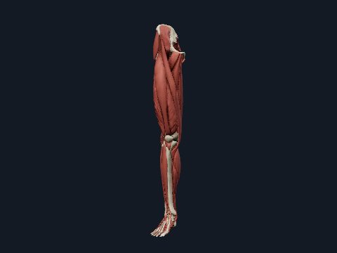
The nerves of the right lower limb together with the muscles they supply.
Open this view in 3DImage: Cogni Anatomy · CC BY-SA 4.0 · Sources: BodyParts3D, Z-Anatomy
What is in this model
90 3D parts. Names follow the Foundational Model of Anatomy (FMA).
- Muscles 58
- Nerves 32
- Abductor digiti minimi of right foot
- Abductor hallucis
- Adductor brevis
- Adductor longus
- Adductor magnus
- Adductor minimus
- Anterior branch of obturator nerve
- Anterior cutaneous branches of femoral nerve
- Common fibular nerve
- Common plantar digital branches of lateral plantar nerve
- Common plantar digital branches of medial plantar nerve
- Deep fibular nerve
- Dorsal digital branches of deep fibular nerve
- Dorsal digital branches of superficial fibular nerve
- Extensor digitorum longus
- Extensor hallucis brevis
- Extensor hallucis longus
- Femoral branch of genitofemoral nerve
- Femoral nerve
- Fibularis brevis
- Fibularis longus
- Fibularis tertius
- First lumbrical of right foot
- First plantar interosseous of right foot
- Flexor accessorius
- Flexor digiti minimi brevis of right foot
- Flexor digitorum brevis
- Flexor digitorum longus
- Flexor hallucis longus
- Fourth lumbrical of right foot
- Gemellus inferior
- Gemellus superior
- Gluteus maximus
- Gluteus medius
- Gluteus minimus
- Gracilis
- Iliacus
- Infrapatellar branch of saphenous nerve
- Intermediate dorsal cutaneous nerve of foot
- Lateral femoral cutaneous nerve
- and 50 more
Questions
What is the largest nerve in the body?
The sciatic nerve. It forms from the L4–S3 roots of the sacral plexus, leaves the pelvis through the greater sciatic foramen and runs down the back of the thigh.
Which nerve supplies the quadriceps?
The femoral nerve (L2–L4). It also supplies sartorius, iliacus and pectineus, and the skin of the front of the thigh and, through the saphenous nerve, of the medial leg.
What does the common fibular nerve supply?
Through its deep branch, the muscles of the anterior compartment of the leg and the short extensors on the dorsum of the foot; through its superficial branch, fibularis longus and brevis and most of the skin on the top of the foot.