Bones of the leg in 3D
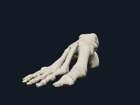
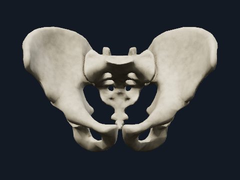
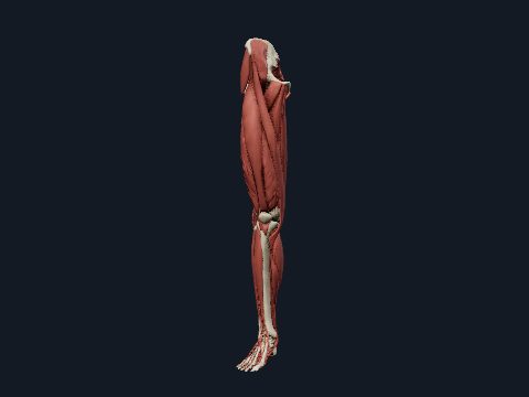
The bones of the leg (lower limb) are the hip bone, the femur of the thigh, the patella, the tibia and fibula of the leg, and the 26 bones of the foot. Built to carry body weight in standing and walking, they form the hip, knee and ankle joints. This free 3D model shows the skeleton of the right lower limb, which you can rotate, take apart and study bone by bone.
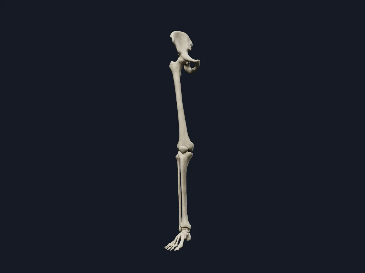 Image: Cogni Anatomy · CC BY 4.0 · Sources: BodyParts3D
- 31 bones in each lower limb, counting the hip bone
- The femur is the longest and strongest bone in the body
- The patella is the largest sesamoid bone, set in the quadriceps tendon
- The tibia carries the body's weight in the leg; the fibula carries little and mainly anchors muscles
- 26 bones in each foot: 7 tarsals, 5 metatarsals and 14 phalanges
Structure of the lower limb skeleton
The hip bone forms from the ilium, ischium and pubis, which fuse at the acetabulum. The head of the femur sits deep in this socket, so the hip joint is far more stable than the shoulder. The femoral neck angles the shaft inward, bringing the knees close to the midline under the body's centre of gravity.
At the knee the femoral condyles rest on the flat tibial plateau, and the patella glides in front of the femur in the patellar groove. The fibula takes no part in the knee joint. At the ankle the tibia and fibula grip the talus between the medial and lateral malleoli.
What the 3D model shows
The model shows the bones of the right lower limb: the hip bone, femur, patella, tibia and fibula, the seven tarsal bones (talus, calcaneus, navicular, cuboid and three cuneiforms), the five metatarsals, the phalanges of the toes and the sesamoid bones under the first metatarsal. The bones are shown on their own, so the surfaces of the hip, knee and ankle joints can be seen from any angle, and the explode slider separates them to show each articular surface.
How to study the leg bones
Start in the anterior view and follow the femur from its head in the acetabulum to the condyles at the knee. Rotate to the lateral view to see that the fibula lies behind and to the side of the tibia and reaches lower, ending as the lateral malleolus. Isolate the femur and find the greater and lesser trochanters, the linea aspera on its posterior surface and the adductor tubercle. Use the section tool through the knee to see how the patella sits in front of the femur, then name the bones from hip to toe from memory.
What is in this model
33 3D parts. Names follow the Foundational Model of Anatomy (FMA).
- Bones 33
- Calcaneus
- Cuboid bone
- Distal phalanx of right big toe
- Distal phalanx of right fourth toe
- Distal phalanx of right little toe
- Distal phalanx of right second toe
- Distal phalanx of right third toe
- Femur
- Fibula
- Fifth metatarsal bone
- First metatarsal bone
- Fourth metatarsal bone
- Hip bone
- Intermediate cuneiform bone
- Lateral cuneiform bone
- Medial cuneiform bone
- Middle phalanx of right fourth toe
- Middle phalanx of right little toe
- Middle phalanx of right second toe
- Middle phalanx of right third toe
- Navicular bone of right foot
- Patella
- Proximal phalanx of right big toe
- Proximal phalanx of right fourth toe
- Proximal phalanx of right little toe
- Proximal phalanx of right second toe
- Proximal phalanx of right third toe
- Second metatarsal bone
- Sesamoid bone of right foot
- Talus
- Third metatarsal bone
- Tibia
Questions
How many bones are in the leg?
Each lower limb has 31 bones: the hip bone, femur, patella, tibia, fibula and the 26 bones of the foot. In the strict anatomical sense, the leg between knee and ankle has two bones, the tibia and the fibula.
What is the strongest bone in the body?
The femur, which is also the longest. Its length is roughly a quarter of a person's height.
Is the kneecap a bone?
Yes. The patella is a sesamoid bone that develops in the tendon of quadriceps femoris and increases the leverage of the quadriceps at the knee.