Muscles of the leg in 3D
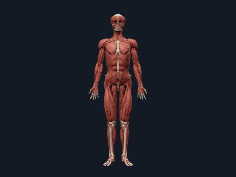
The muscles of the leg (lower limb) steady the pelvis, move the hip, knee, ankle and toes, and keep the body upright in standing and walking. The thigh has anterior, medial and posterior compartments, the leg has anterior, lateral and posterior ones, and the sole has four layers of intrinsic muscles. This free 3D model shows the muscles of the right lower limb on its skeleton, layer by layer.
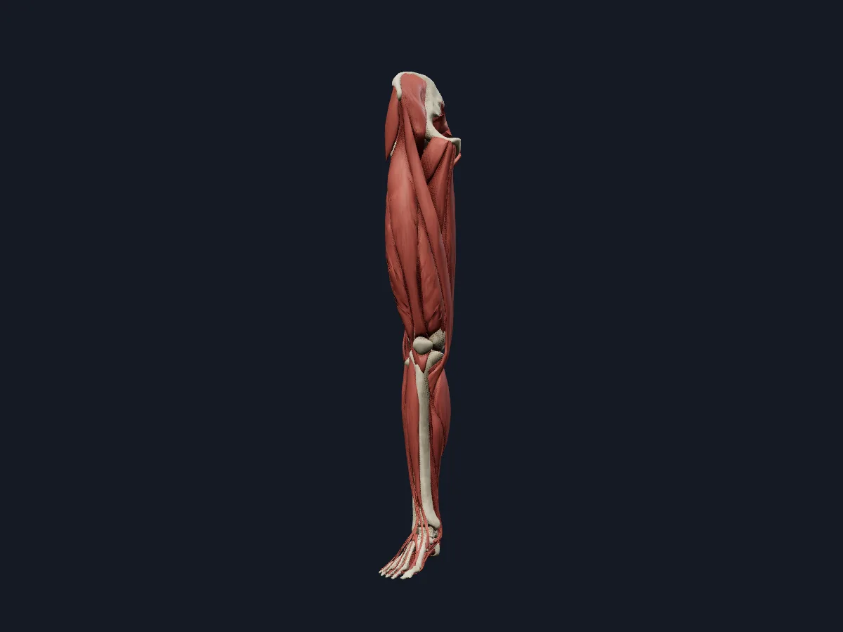 Image: Cogni Anatomy · CC BY 4.0 · Sources: BodyParts3D
- Gluteus maximus is the largest muscle in the body
- Sartorius is the longest muscle in the body
- Quadriceps femoris has 4 parts: rectus femoris, vastus lateralis, vastus intermedius and vastus medialis
- 3 hamstrings: biceps femoris, semitendinosus and semimembranosus
- Gastrocnemius and soleus insert together through the calcaneal (Achilles) tendon, the strongest tendon in the body
Muscle groups of the lower limb
The gluteal muscles extend and abduct the hip, and the deep lateral rotators (piriformis, the gemelli, obturator internus and externus, quadratus femoris) turn the femur outward. In the thigh, the quadriceps in the anterior compartment extends the knee and is supplied by the femoral nerve; the adductors in the medial compartment are supplied mainly by the obturator nerve; the hamstrings at the back extend the hip, flex the knee and are supplied by the sciatic nerve.
Below the knee, the anterior compartment dorsiflexes the foot, the fibularis muscles of the lateral compartment evert it, and the calf muscles of the posterior compartment plantarflex it.
What the 3D model shows
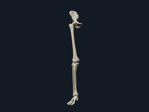
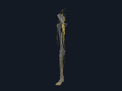
The model shows the muscles of the right lower limb on its bones: gluteus maximus, medius and minimus, tensor fasciae latae and the deep rotators; iliacus, sartorius, the quadriceps and the adductor group with gracilis and pectineus; the hamstrings; the anterior, lateral and posterior leg muscles, including plantaris and popliteus; and the intrinsic muscles of the foot. The iliotibial tract and the calcaneal tendon are included. A second view adds the arteries, veins and nerves of the limb, along with some neighbouring pelvic structures.
How to study the leg muscles
Use the posterior view first: find gluteus maximus, then go to the deeper muscle layers to expose gluteus medius and the deep rotators below piriformis. In the anterior view, trace sartorius from the anterior superior iliac spine across the thigh to the medial side of the knee, and name the four parts of the quadriceps. Then isolate the calf: hide gastrocnemius to see soleus and follow both into the calcaneal tendon. Finish by listing each compartment's muscles and nerve from memory and checking them in the model.
Views in the 3D model
The muscles of the right lower limb on its bones, from the gluteal region to the sole of the foot.
Open this view in 3DImage: Cogni Anatomy · CC BY 4.0 · Sources: BodyParts3D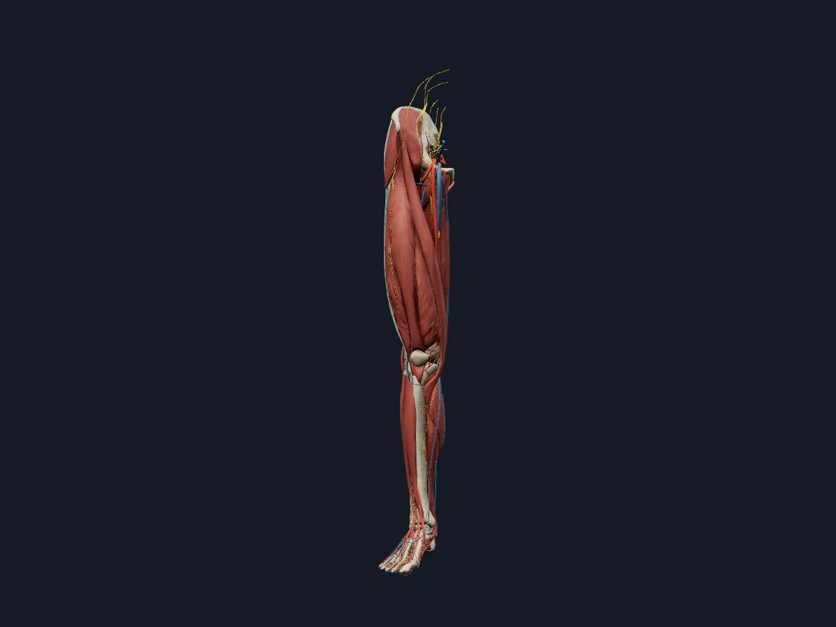
Every layer of the right lower limb together: bones, muscles, arteries, veins and nerves, with neighbouring pelvic structures.
Open this view in 3DImage: Cogni Anatomy · CC BY-SA 4.0 · Sources: BodyParts3D, Z-Anatomy
What is in this model
189 3D parts. Names follow the Foundational Model of Anatomy (FMA).
- Muscles 58
- Bones 33
- Nerves 32
- Arteries 31
- Veins 31
- Connective tissue 4
- Abductor digiti minimi of right foot
- Abductor hallucis
- Adductor brevis
- Adductor longus
- Adductor magnus
- Adductor minimus
- Anterior branch of obturator nerve
- Anterior cutaneous branches of femoral nerve
- Anterior tibial artery
- Anterior tibial recurrent artery
- Anterior tibial vein
- Arcuate artery
- Calcaneal tendon
- Calcaneus
- Common fibular nerve
- Common plantar digital branches of lateral plantar nerve
- Common plantar digital branches of medial plantar nerve
- Cuboid bone
- Deep femoral vein
- Deep fibular nerve
- Deep plantar artery
- Descending branch of right lateral circumflex femoral artery
- Descending genicular artery
- Distal perforating artery
- Distal phalanx of right big toe
- Distal phalanx of right fourth toe
- Distal phalanx of right little toe
- Distal phalanx of right second toe
- Distal phalanx of right third toe
- Dorsal digital artery of foot
- Dorsal digital branches of deep fibular nerve
- Dorsal digital branches of superficial fibular nerve
- Dorsal metacarpal vein
- Dorsal venous arch of right foot
- Dorsalis pedis artery
- Extensor digitorum longus
- Extensor hallucis brevis
- Extensor hallucis longus
- Femoral artery
- Femoral branch of genitofemoral nerve
- and 137 more
Questions
What are the main muscles of the leg?
In the thigh: the quadriceps at the front, the hamstrings at the back and the adductors on the inner side, with the gluteal muscles above them at the hip. Below the knee: tibialis anterior and the toe extensors at the front, the fibularis muscles on the outer side, and gastrocnemius and soleus in the calf.
Which muscles form the calf?
Gastrocnemius, with its medial and lateral heads, and soleus beneath it, together called triceps surae. Both insert on the calcaneus through the calcaneal (Achilles) tendon; the small plantaris lies between them.
What is the largest muscle in the human body?
Gluteus maximus, which forms most of the buttock and extends and laterally rotates the hip. The longest muscle, sartorius, is also in the lower limb.