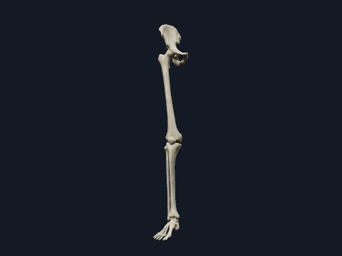
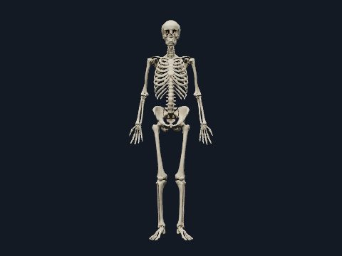
Bones of the foot in 3D
The foot skeleton has 26 bones: 7 tarsal bones in the hindfoot and midfoot, 5 metatarsals and 14 phalanges in the toes. They are arranged in longitudinal and transverse arches that spread body weight between the heel and the ball of the foot. In this free 3D model you can rotate the right foot, take it apart and study its bones from every side.
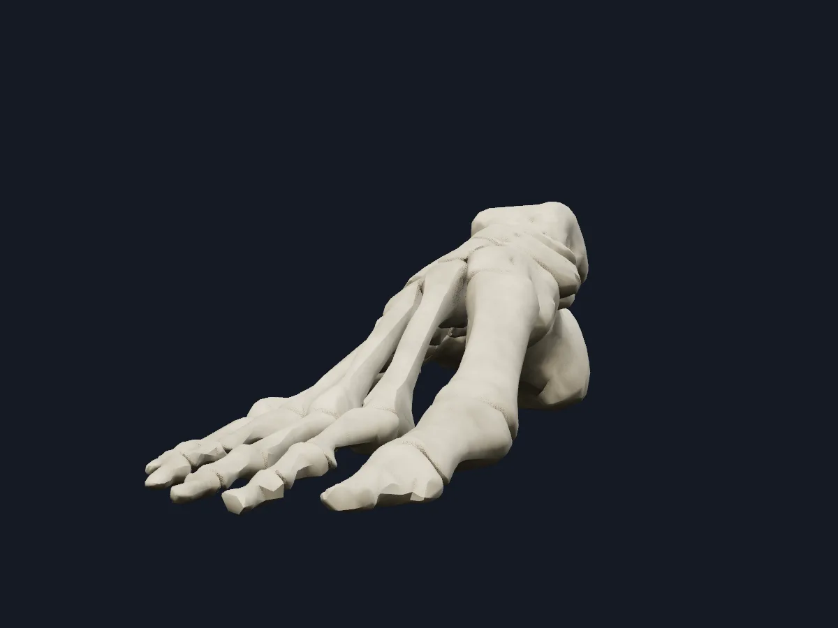 Image: Cogni Anatomy · CC BY 4.0 · Sources: BodyParts3D
- 26 bones in each foot: 7 tarsals, 5 metatarsals and 14 phalanges
- The calcaneus is the largest tarsal bone and forms the heel
- The talus links the leg to the foot and has no muscles attached to it
- The big toe (hallux) has 2 phalanges; each of the other toes has 3
- 3 arches: medial longitudinal, lateral longitudinal and transverse
Structure of the foot skeleton
The hindfoot consists of the talus and calcaneus. The talus sits in the ankle mortise formed by the tibia and fibula and rests on the calcaneus at the subtalar joint. The midfoot holds the navicular, the cuboid and the three cuneiforms; the forefoot holds the metatarsals and phalanges.
The medial longitudinal arch, higher than the lateral, runs from the calcaneus through the talus, navicular and cuneiforms to the first three metatarsals. The lateral arch passes through the calcaneus, cuboid and the fourth and fifth metatarsals. The first metatarsal is the shortest and thickest.
What the 3D model shows
The model shows the bones of the right foot on their own: the talus, calcaneus, navicular, cuboid and the medial, intermediate and lateral cuneiforms, the five metatarsals, the proximal, middle and distal phalanges of the toes, and the two sesamoid bones under the head of the first metatarsal. Without soft tissue, the joint surfaces of the tarsus and the shape of the arches are easy to see from the medial and inferior views. The explode slider separates every bone so you can examine its articular facets.
How to study the foot bones
Start from the superior (dorsal) view and name the bones in rows: talus and calcaneus at the back, then the midfoot, then the metatarsals numbered from the big toe. Rotate to the medial view to see the medial longitudinal arch, and to the inferior view for the underside of the calcaneus. Isolate the talus and find its trochlea, neck and head. Then explode the foot, pick single bones and identify them without the surrounding skeleton.
What is in this model
28 3D parts. Names follow the Foundational Model of Anatomy (FMA).
- Bones 28
- Calcaneus
- Cuboid bone
- Distal phalanx of right big toe
- Distal phalanx of right fourth toe
- Distal phalanx of right little toe
- Distal phalanx of right second toe
- Distal phalanx of right third toe
- Fifth metatarsal bone
- First metatarsal bone
- Fourth metatarsal bone
- Intermediate cuneiform bone
- Lateral cuneiform bone
- Medial cuneiform bone
- Middle phalanx of right fourth toe
- Middle phalanx of right little toe
- Middle phalanx of right second toe
- Middle phalanx of right third toe
- Navicular bone of right foot
- Proximal phalanx of right big toe
- Proximal phalanx of right fourth toe
- Proximal phalanx of right little toe
- Proximal phalanx of right second toe
- Proximal phalanx of right third toe
- Second metatarsal bone
- Sesamoid bone of right foot
- Talus
- Third metatarsal bone
Questions
How many bones are in the foot?
26: 7 tarsals, 5 metatarsals and 14 phalanges. The two feet together have 52 bones, about a quarter of the bones in the adult body.
What are the 7 tarsal bones?
The talus, calcaneus, navicular, cuboid and the medial, intermediate and lateral cuneiforms.
Is this 3D foot model free?
Yes. The foot model and the rest of the atlas are free to use in the browser, without an account.