Brain ventricles in 3D
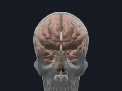
The ventricles of the brain are four connected cavities filled with cerebrospinal fluid (CSF): two lateral ventricles, the third ventricle and the fourth ventricle. CSF is produced mainly by the choroid plexus and flows from the lateral ventricles through the interventricular foramina, the third ventricle and the cerebral aqueduct into the fourth ventricle and out to the subarachnoid space. In this free 3D model the ventricular system is shown inside a see-through brain, so you can turn it and follow the path of CSF.
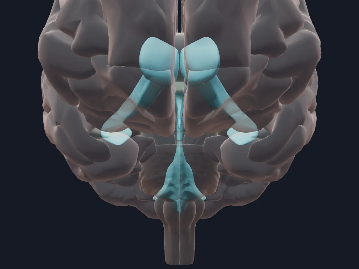 Image: Cogni Anatomy · CC BY 4.0 · Sources: BodyParts3D
- 4 ventricles: 2 lateral, the third and the fourth
- Each lateral ventricle has a central part and anterior, posterior and inferior horns
- About 150 mL of CSF in adults, produced at roughly 500 mL per day
- The fourth ventricle opens into the subarachnoid space through one median and two lateral apertures
- Below the fourth ventricle the system continues as the central canal of the spinal cord
Structure of the ventricular system
Each lateral ventricle is a C-shaped cavity in a cerebral hemisphere: the anterior horn lies in the frontal lobe, the central part under the corpus callosum, the posterior horn in the occipital lobe and the inferior horn in the temporal lobe. Through the interventricular foramen (of Monro) each opens into the third ventricle, a narrow midline slit between the two thalami.
The cerebral aqueduct (of Sylvius) runs through the midbrain and connects the third ventricle with the fourth, a tent-shaped cavity between the pons and medulla in front and the cerebellum behind.
What the 3D model shows
The view shows the ventricular system as a cast of six parts: the left and right lateral ventricles, the interventricular foramen, the third ventricle, the cerebral aqueduct and the fourth ventricle. The rest of the brain is faded around it, so you can see that the anterior horns lie in the frontal lobes, the inferior horns curve down into the temporal lobes, and the fourth ventricle sits in front of the cerebellum. Each cavity can be selected, hidden or isolated.
How to study the ventricles
Begin with the lateral view to see the C shape of a lateral ventricle and name its horns. Turn to the superior view, where the two lateral ventricles lie side by side, and then to the anterior view to find the interventricular foramina leading into the slit-like third ventricle. From the lateral view again, follow the cerebral aqueduct down to the fourth ventricle. Trace one drop of CSF from a lateral ventricle to the subarachnoid space, then repeat the route aloud with the model closed.
What is in this model
6 3D parts. Names follow the Foundational Model of Anatomy (FMA).
- Nerves 6
- Cerebral aqueduct
- Fourth ventricle
- Interventricular foramen
- Lateral ventricle
- Third ventricle
Questions
How many ventricles are in the brain?
Four: the right and left lateral ventricles in the cerebral hemispheres, the third ventricle in the diencephalon and the fourth ventricle between the brainstem and the cerebellum.
Where is cerebrospinal fluid produced?
Mainly by the choroid plexus in the lateral, third and fourth ventricles. It leaves the ventricular system through the apertures of the fourth ventricle into the subarachnoid space.
What connects the third and fourth ventricles?
The cerebral aqueduct, a narrow channel that runs through the midbrain.