Brain anatomy in 3D
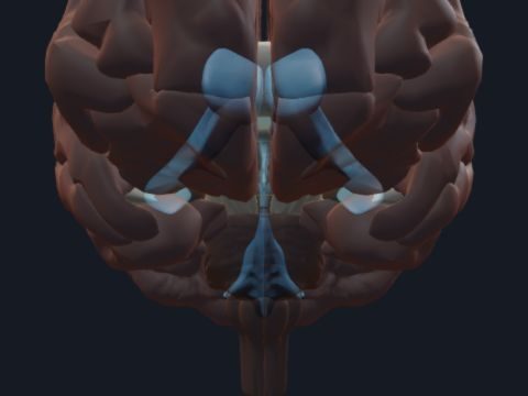
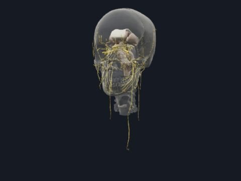
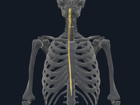
The brain is the part of the central nervous system inside the skull, made up of the cerebrum, diencephalon, brainstem and cerebellum. Its folded cortex and deep nuclei process sensation, movement, memory and language, and the brainstem connects it with the spinal cord. In this free 3D model you can turn the brain inside the skull or on its own, cut through it to reach the deep structures, and isolate any gyrus or nucleus by name.
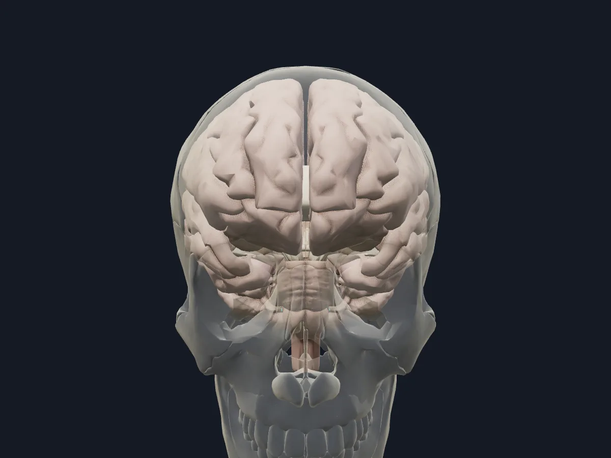 Image: Cogni Anatomy · CC BY 4.0 · Sources: BodyParts3D
- Weighs about 1.3–1.4 kg in adults
- Contains about 86 billion neurons
- Each hemisphere has frontal, parietal, temporal and occipital lobes, plus the insula hidden in the lateral sulcus
- The central sulcus separates the precentral gyrus (primary motor cortex) from the postcentral gyrus (primary somatosensory cortex)
- The corpus callosum is the largest commissure connecting the two hemispheres
Parts of the brain
The cerebrum consists of two hemispheres with an outer cortex folded into gyri and sulci, underlying white matter and deep grey nuclei. The putamen and globus pallidus form the lentiform nucleus. The anterior limb of the internal capsule separates it from the head of the caudate nucleus; the posterior limb separates it from the thalamus. The cingulate and parahippocampal gyri lie on the medial surface. The hippocampus and amygdala lie in the medial temporal lobe. The fornix carries major connections of the hippocampal formation, including projections to the mammillary bodies.
The diencephalon contains the thalamus, the hypothalamus with the mammillary bodies, and the pineal body. The brainstem is formed by the midbrain, pons and medulla oblongata, and the cerebellum sits behind it in the posterior cranial fossa.
What the 3D model shows
The model has 107 parts. On the surface you can identify the superior, middle and inferior frontal gyri, the precentral and postcentral gyri, the superior parietal lobule, supramarginal and angular gyri, the superior, middle and inferior temporal gyri, the orbital gyri, the occipital lobe and the insula. Inside are the white matter of each hemisphere, the corpus callosum, fornix and commissures, the caudate nucleus, putamen, globus pallidus and internal capsule, the thalamus, hypothalamus and geniculate bodies, the hippocampus and amygdala, the ventricles with the choroid plexus, and the midbrain, pons, medulla oblongata and cerebellum.
How to study the brain
Start in the isolated view from the side: find the lateral and central sulci and name the gyri of each lobe. Make the white matter see-through or hide one hemisphere to look at the medial surface, the corpus callosum and the thalamus. Then open the section view and compare the caudate nucleus, putamen and globus pallidus on either side of the internal capsule. Finish in the brain-in-skull view, matching the frontal lobe, temporal lobe and cerebellum to the cranial fossae. A day later, sketch a lateral and a medial view from memory.
Views in the 3D model
The brain in place inside a faded skull, showing how it fills the cranial cavity.
Open this view in 3DImage: Cogni Anatomy · CC BY 4.0 · Sources: BodyParts3D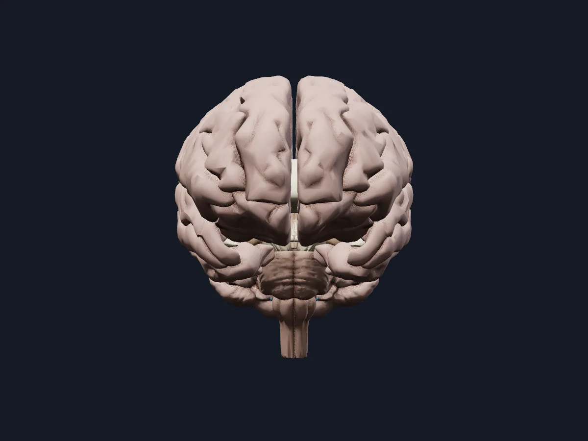
The brain on its own, with the cerebral hemispheres, brainstem and cerebellum.
Open this view in 3DImage: Cogni Anatomy · CC BY 4.0 · Sources: BodyParts3D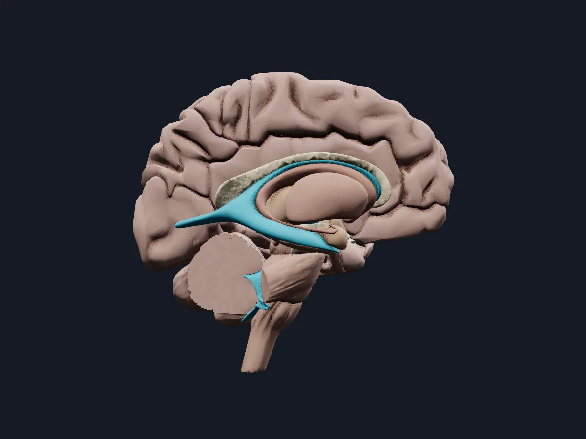
The brain cut open to show the basal nuclei, thalamus, limbic structures and ventricles.
Open this view in 3DImage: Cogni Anatomy · CC BY 4.0 · Sources: BodyParts3D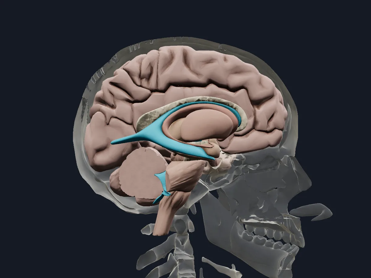
A section through the brain with the skull faded around it.
Open this view in 3DImage: Cogni Anatomy · CC BY 4.0 · Sources: BodyParts3D
What is in this model
107 3D parts. Names follow the Foundational Model of Anatomy (FMA).
- Nerves 106
- Endocrine 1
- Amygdala
- Angular gyrus
- Anterior commissure
- Anterior part of left superior temporal gyrus
- Anterior part of right superior temporal gyrus
- Brachium of left inferior colliculus
- Brachium of left superior colliculus
- Brachium of right inferior colliculus
- Brachium of right superior colliculus
- Caudate nucleus
- Cerebellum
- Cerebral aqueduct
- Choroid plexus of cerebral hemisphere
- Cingulate gyrus
- Commissure of fornix of forebrain
- Corpus callosum
- Fornix of forebrain
- Fourth ventricle
- Fusiform gyrus
- Globus pallidus
- Habenula
- Hippocampus
- Hypothalamus
- Inferior colliculus
- Inferior frontal gyrus
- Inferior temporal gyrus
- Insula
- Internal capsule
- Interventricular foramen
- Lamina terminalis
- Lateral geniculate body
- Lateral ventricle
- Mammillary body
- Medial geniculate body
- Medulla oblongata
- Midbrain
- Middle frontal gyrus
- Middle temporal gyrus
- Occipital lobe
- Optic chiasm
- and 24 more
Questions
What are the main parts of the brain?
The cerebrum, made of two hemispheres, the diencephalon with the thalamus and hypothalamus, the brainstem (midbrain, pons and medulla oblongata) and the cerebellum.
How much does the human brain weigh?
About 1.3–1.4 kg in an adult, roughly 2 % of body weight.
Where is the hippocampus?
In the medial part of the temporal lobe, in the floor of the inferior horn of the lateral ventricle. It continues backwards into the fornix.