Spinal cord anatomy in 3D
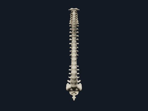
The spinal cord is the part of the central nervous system inside the vertebral canal. It continues from the medulla oblongata at the foramen magnum and ends at about the level of the first and second lumbar vertebrae, giving off a pair of spinal nerves from each segment. In this free 3D model you can see the cord segment by segment from C1 to S5 inside a see-through vertebral column and compare each segment with the vertebrae around it.
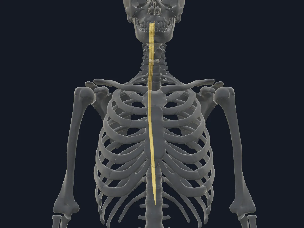 Image: Cogni Anatomy · CC BY 4.0 · Sources: BodyParts3D, HRA
- 31 segments: 8 cervical, 12 thoracic, 5 lumbar, 5 sacral and 1 coccygeal
- 31 pairs of spinal nerves, one pair per segment
- About 42–45 cm long in adults
- Ends as the conus medullaris at about the L1–L2 vertebral level; below it the nerve roots form the cauda equina
- Two enlargements, cervical and lumbosacral, give rise to the nerves of the limbs
Structure of the spinal cord
In cross-section the cord has central H-shaped grey matter with anterior and posterior horns, and a lateral horn in the thoracic and upper lumbar segments, surrounded by white matter in anterior, lateral and posterior funiculi. A narrow central canal runs through the middle. The cord is covered by the dura mater, arachnoid mater and pia mater.
Because the cord is shorter than the vertebral column, segments lie higher than the vertebrae of the same number: the lumbar and sacral segments sit opposite the lower thoracic and first lumbar vertebrae, and their nerve roots run down to reach their foramina.
What the 3D model shows
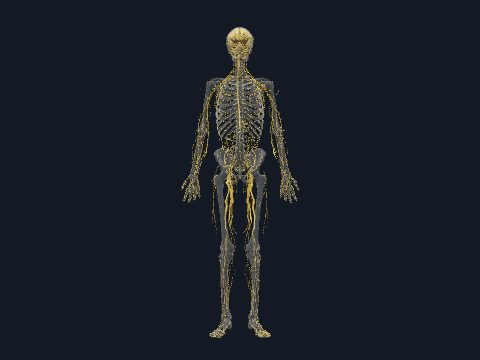
The view shows the spinal cord as 30 separate segments: C1–C8, T1–T12, L1–L5 and S1–S5, plus the central canal, with the skeleton faded around them. Each segment can be selected, so you can see exactly where a given segment lies in relation to the vertebral bodies and how the cord widens in the cervical and lumbosacral regions and narrows towards its end. The view shows the cord itself; spinal nerve roots and the cauda equina appear in the nervous system views.
How to study the spinal cord
Use the lateral view and slide down the cord from C1, noting which vertebra lies beside each group of segments. Isolate the lumbar and sacral segments to see how short and high the lower cord is compared with the lumbar spine. Switch to the posterior view to compare the width of the cervical and lumbosacral enlargements with the thoracic cord. Then test yourself: which vertebral level lies over the L1 segment, and why is that important for the nerve roots below?
What is in this model
31 3D parts. Names follow the Foundational Model of Anatomy (FMA).
- Nerves 31
- C1 segment of cervical spinal cord
- C2 segment of cervical spinal cord
- C3 segment of cervical spinal cord
- C4 segment of cervical spinal cord
- C5 segment of cervical spinal cord
- C6 segment of cervical spinal cord
- C7 segment of cervical spinal cord
- C8 segment of cervical spinal cord
- Central canal of spinal cord
- Eighth thoracic spinal cord segment
- Eleventh thoracic spinal cord segment
- Fifth lumbar spinal cord segment
- Fifth sacral spinal cord segment
- Fifth thoracic spinal cord segment
- First lumbar spinal cord segment
- First sacral spinal cord segment
- First thoracic spinal cord segment
- Fourth lumbar spinal cord segment
- Fourth sacral spinal cord segment
- Fourth thoracic spinal cord segment
- Ninth thoracic spinal cord segment
- Second lumbar spinal cord segment
- Second sacral spinal cord segment
- Second thoracic spinal cord segment
- Seventh thoracic spinal cord segment
- Sixth thoracic spinal cord segment
- Tenth thoracic spinal cord segment
- Third lumbar spinal cord segment
- Third sacral spinal cord segment
- Third thoracic spinal cord segment
- Twelfth thoracic spinal cord segment
Questions
How long is the spinal cord?
About 42–45 cm in adults, much shorter than the vertebral column, which is around 70 cm.
Where does the spinal cord end?
In adults it usually ends as the conus medullaris at about the level of the L1–L2 intervertebral disc. Below this the vertebral canal contains the nerve roots of the cauda equina.
How many spinal nerves are there?
31 pairs: 8 cervical, 12 thoracic, 5 lumbar, 5 sacral and 1 coccygeal, one pair from each segment of the cord.