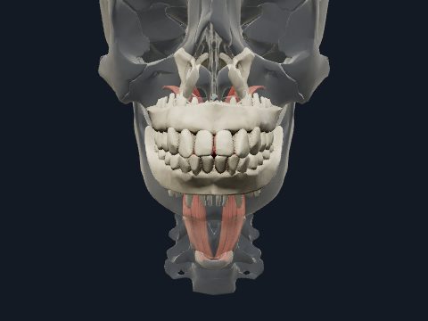
Digestive system anatomy in 3D
The digestive system is the alimentary canal from the mouth to the anus together with the accessory organs, the salivary glands, liver, gallbladder and pancreas, which add secretions to it. It breaks food down, absorbs nutrients and water, and removes the rest. In this free 3D model you can follow the tract from the oesophagus to the rectum and see the liver segments, bile ducts and pancreatic duct.
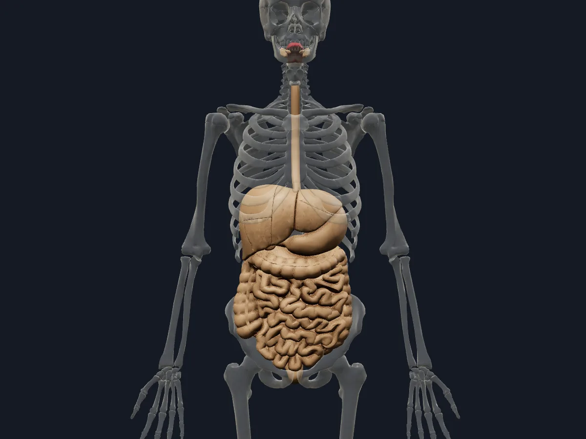 Image: Cogni Anatomy · CC BY 4.0 · Sources: BodyParts3D
- The oesophagus is about 25 cm long, from the pharynx at C6 to the stomach at about T11
- The small intestine is about 6–7 m long: duodenum (about 25 cm), jejunum and ileum
- The large intestine is about 1.5 m long, and the colon carries 3 bands of longitudinal muscle, the taeniae coli
- The liver is the largest gland in the body, about 1.5 kg in adults
- 3 pairs of major salivary glands: parotid, submandibular and sublingual
Parts of the digestive system
Food passes from the mouth through the pharynx and oesophagus into the stomach, then through the C-shaped duodenum, the jejunum and the ileum. At the ileocaecal junction it enters the large intestine: caecum with the appendix, ascending, transverse, descending and sigmoid colon, rectum and anal canal.
The liver lies under the right dome of the diaphragm and divides into eight functional segments, each with its own branches of the portal vein, hepatic artery and bile duct. Bile drains through the hepatic ducts and is stored in the gallbladder; the pancreas sits behind the stomach with its head in the curve of the duodenum.
What the 3D model shows
The model has 106 digestive parts: the tongue and the submandibular and sublingual glands; the oesophagus, stomach and duodenum; the jejunum and ileum in proximal, middle and distal parts with their mesentery; the ileocaecal junction, appendix and mesoappendix; the ascending, transverse and descending colon with the taeniae and transverse mesocolon; and the rectum. The liver is divided into its segments with the caudate lobe, intrahepatic bile tributaries, hepatic, common hepatic and cystic ducts, and gallbladder; the pancreas shows its parenchyma and duct tree.
How to study the digestive system
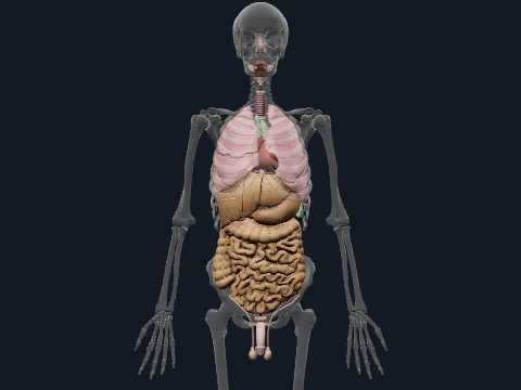
Begin with the system inside the see-through skeleton in the anterior view to place each organ in its abdominal region. Switch to the isolated view and turn to the posterior view to see the duodenum curve around the pancreas. Use the explode slider to separate the liver segments and number them. Trace bile from the intrahepatic tributaries through the hepatic ducts to the gallbladder, then trace food from the oesophagus to the rectum. Close the model and repeat both routes from memory.
Views in the 3D model
The digestive organs in place inside a see-through skeleton, from the tongue and salivary glands to the rectum.
Open this view in 3DImage: Cogni Anatomy · CC BY 4.0 · Sources: BodyParts3D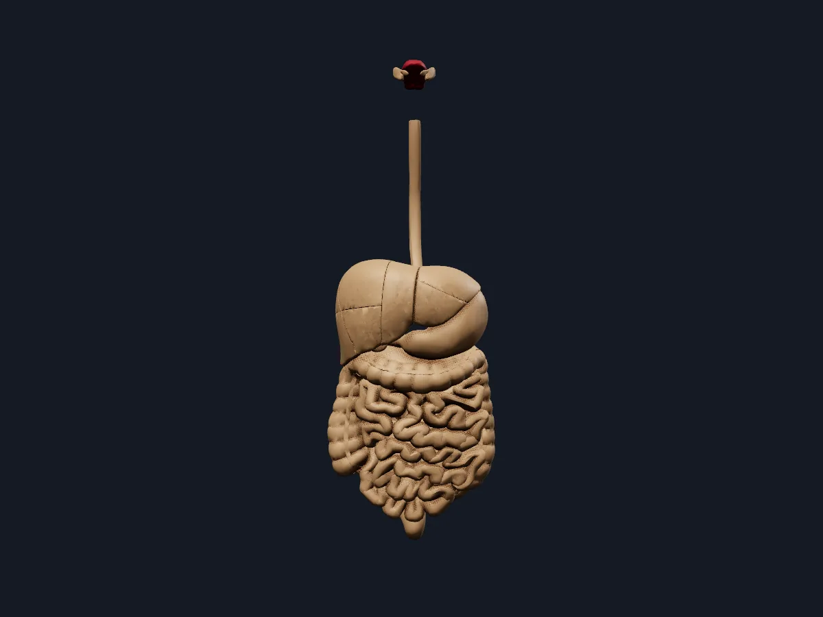
The digestive tract, liver, gallbladder and pancreas on their own, without surrounding structures.
Open this view in 3DImage: Cogni Anatomy · CC BY 4.0 · Sources: BodyParts3D
What is in this model
106 3D parts. Names follow the Foundational Model of Anatomy (FMA).
- Digestive 106
- Anterior inferior tributary of right hepatic biliary tree
- Anterior superior tributary of right hepatic biliary tree
- Appendix
- Ascending colon
- Caudate lobe of liver
- Caudate lobe tributary of left hepatic biliary tree
- Common hepatic duct
- Cystic duct
- Descending colon
- Distal part of ileum
- Distal part of jejunum
- Duct of caudate lobe of liver
- Duodenum
- Esophagus
- Gallbladder
- Hepatic duct
- Hepatovenous segment II
- Hepatovenous segment III
- Hepatovenous segment IV
- Hepatovenous segment IX
- Hepatovenous segment V
- Hepatovenous segment VI
- Hepatovenous segment VII
- Hepatovenous segment VIII
- Ileocecal junction
- Lateral inferior tributary of left hepatic biliary tree
- Lateral superior tributary of left hepatic biliary tree
- Medial inferior tributary of left hepatic biliary tree
- Medial superior tributary of left hepatic biliary tree
- Mesentery of small intestine
- Mesoappendix
- Middle part of ileum
- Middle part of jejunum
- Pancreas
- Pancreatic duct
- Pancreatic duct tree
- Parenchyma of pancreas
- Posterior inferior tributary of right hepatic biliary tree
- Posterior superior tributary of right hepatic biliary tree
- Proximal part of ileum
- and 11 more
Questions
What organs are in the digestive system?
The alimentary canal (mouth, pharynx, oesophagus, stomach, small intestine, large intestine and anal canal) and the accessory organs: tongue, teeth, salivary glands, liver, gallbladder and pancreas.
How long is the small intestine?
About 6–7 m in adults. The duodenum is about 25 cm; of the rest, roughly two-fifths is jejunum and three-fifths ileum.
What are the segments of the liver?
In the Couinaud scheme the liver has eight functional segments, numbered I to VIII. Each has its own portal vein, hepatic artery and bile duct branch, and the hepatic veins run between them.