Oral cavity anatomy in 3D
The oral cavity is the first part of the digestive tract, bounded by the lips and cheeks, the palate above and the floor of the mouth below, and opening into the oropharynx behind. It contains the teeth, gums and tongue and is divided into the vestibule and the oral cavity proper. In this free 3D model you can see the upper and lower teeth with the gums, the tongue and the muscles of the soft palate, and isolate any single tooth.
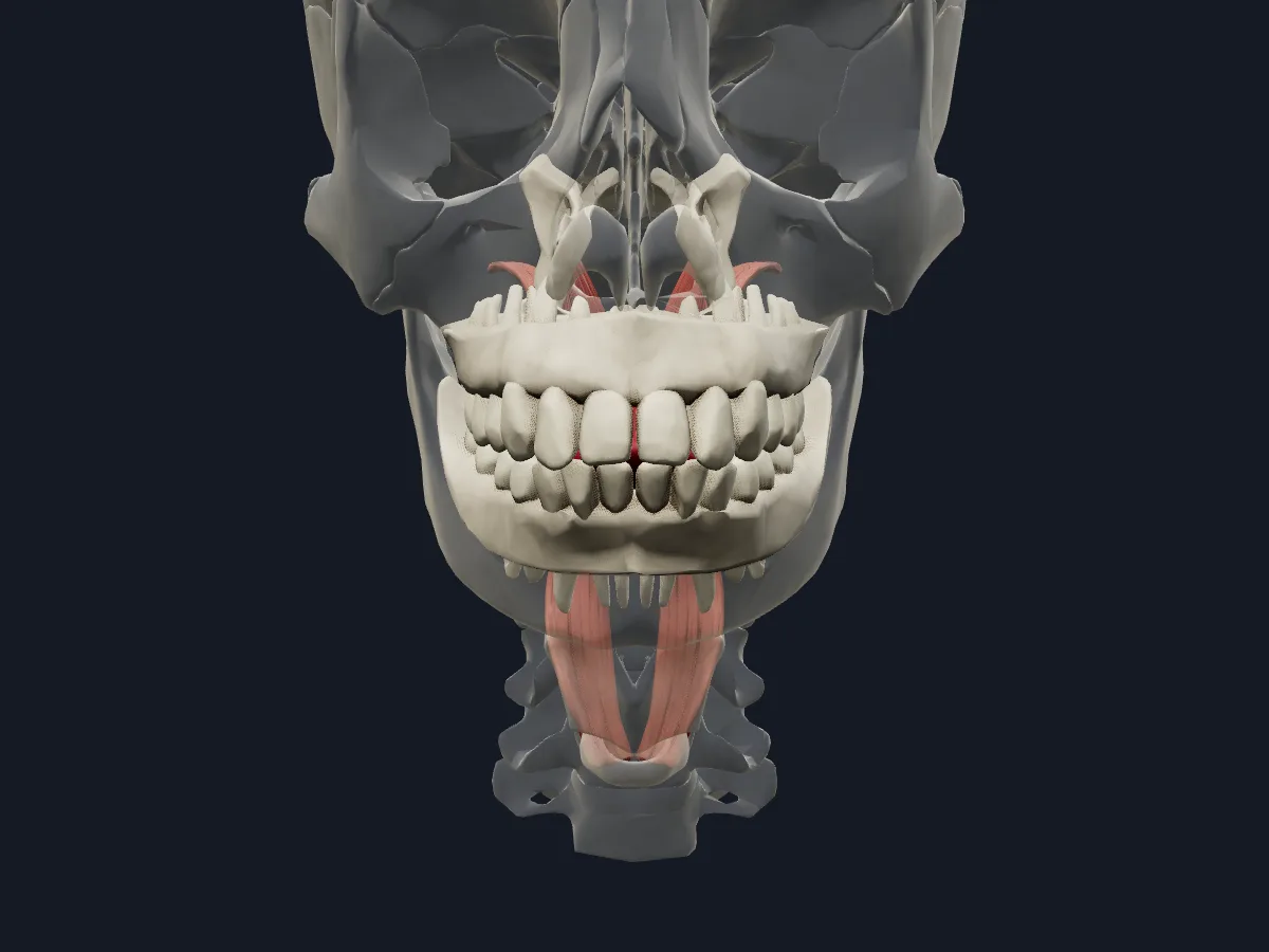 Image: Cogni Anatomy · CC BY 4.0 · Sources: BodyParts3D
- 32 permanent teeth in adults: in each quadrant 2 incisors, 1 canine, 2 premolars and 3 molars
- 20 deciduous (milk) teeth in children
- The hard palate is formed by the palatine processes of the maxillae and the horizontal plates of the palatine bones
- 5 muscles of the soft palate: tensor veli palatini, levator veli palatini, palatoglossus, palatopharyngeus and musculus uvulae
- The tensor veli palatini is supplied by the mandibular nerve; the other soft palate muscles by the vagus nerve
Structure of the oral cavity
The oral vestibule is the slit between the lips and cheeks and the teeth and gums. The oral cavity proper lies inside the dental arches, with the hard and soft palate as its roof and the tongue resting on its floor. The teeth sit in the alveolar processes of the maxillae and mandible, covered at their necks by the gingiva.
The tongue has an oral part, the anterior two-thirds, and a pharyngeal part, the posterior third, separated by the sulcus terminalis. The soft palate hangs from the back of the hard palate, ends in the uvula and rises during swallowing to close off the nasopharynx.
What the 3D model shows
The view has 40 parts with the rest of the skeleton faded. It shows 28 permanent teeth, the central and lateral incisors, canine, first and second premolars and first and second molars in each quadrant, without third molars. The upper and lower gingiva, the tongue and the palatine bones are included. At the back of the palate are the levator and tensor veli palatini, the palatopharyngeus and the uvular muscle, so you can see how the soft palate attaches.
How to study the oral cavity
Begin with the anterior view and count the teeth of one quadrant from the midline: incisors, canine, premolars, molars. Turn to the inferior view to look at the upper dental arch from below, and to the superior view for the lower arch. Isolate one tooth of each type and compare the crowns. Then hide the tongue and use the posterior view to follow the soft palate muscles from the palate towards the pharynx. Finish by writing the adult dental formula and the nerve supply of each palate muscle from memory.
What is in this model
40 3D parts. Names follow the Foundational Model of Anatomy (FMA).
- Bones 32
- Muscles 7
- Digestive 1
- Gingiva of lower jaw
- Gingiva of upper jaw
- Levator veli palatini
- Lower central secondary incisor tooth
- Lower first secondary molar tooth
- Lower first secondary premolar tooth
- Lower lateral secondary incisor tooth
- Lower second secondary molar tooth
- Lower second secondary premolar tooth
- Lower secondary canine tooth
- Palatine bone
- Palatopharyngeus
- Tensor veli palatini
- Tongue
- Upper central secondary incisor tooth
- Upper first secondary molar tooth
- Upper first secondary premolar tooth
- Upper lateral secondary incisor tooth
- Upper second secondary molar tooth
- Upper second secondary premolar tooth
- Upper secondary canine tooth
- Uvular muscle
Questions
How many teeth do adults have?
32 permanent teeth: 8 incisors, 4 canines, 8 premolars and 12 molars, including the four third molars (wisdom teeth). Children have 20 deciduous teeth.
What is the difference between the oral vestibule and the oral cavity proper?
The vestibule is the space between the lips and cheeks on the outside and the teeth and gums on the inside. The oral cavity proper is the space inside the dental arches, bounded by the palate and the floor of the mouth.
Which muscles move the soft palate?
The tensor veli palatini and levator veli palatini tense and raise it, the palatoglossus and palatopharyngeus pull it down, and the musculus uvulae shortens the uvula.