Ear anatomy in 3D
The ear is the organ of hearing and balance, divided into the external, middle and inner ear. Sound collected by the auricle travels along the external acoustic meatus to the tympanic membrane, is passed by the three auditory ossicles to the oval window and is converted into nerve signals in the cochlea. In this free 3D model you can see the middle and inner ear inside the temporal bone, enlarge the ossicles and view the external ear on its own.
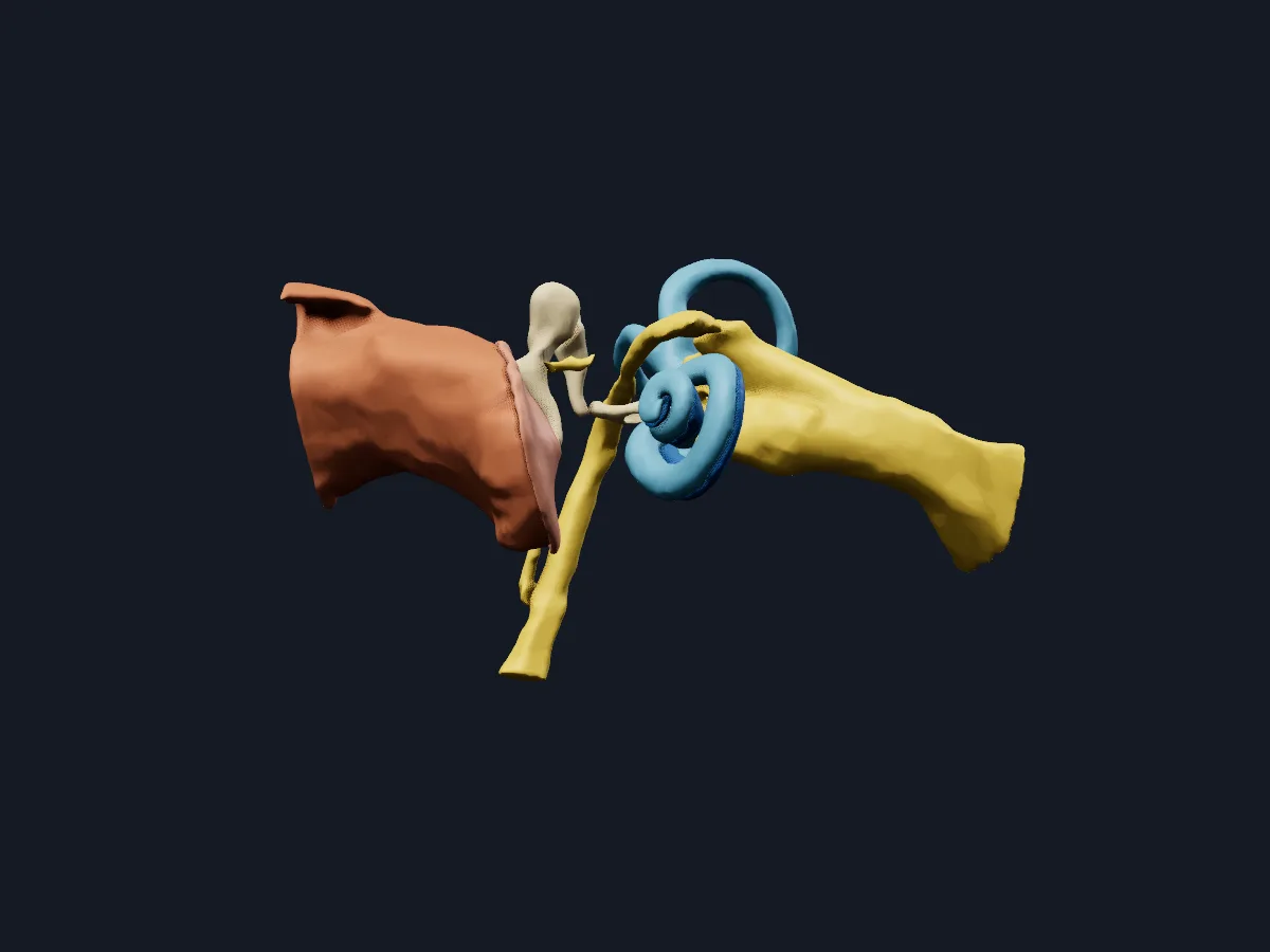 Image: Cogni Anatomy · CC BY 4.0 · Sources: OpenEar
- 3 parts: external, middle and inner ear
- 3 auditory ossicles: malleus, incus and stapes
- The stapes is the smallest bone in the human body, about 3 mm long
- The cochlea makes about two and a half turns
- The vestibulocochlear nerve (CN VIII) has a cochlear part for hearing and a vestibular part for balance
Structure of the ear
The external ear consists of the auricle and the external acoustic meatus, whose outer third is cartilaginous and inner two-thirds bony; the tympanic membrane closes it medially. The middle ear is an air-filled cavity in the temporal bone, connected to the nasopharynx by the pharyngotympanic tube. The handle of the malleus is attached to the tympanic membrane, the incus links it to the stapes, and the footplate of the stapes sits in the oval window.
The inner ear is a bony labyrinth with the vestibule, three semicircular canals and the cochlea. In the cochlea, the scala vestibuli and scala tympani meet at its apex.
What the 3D model shows
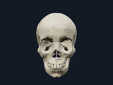
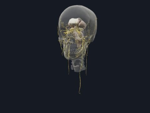
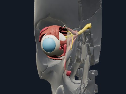
The middle and inner ear view shows the tympanic membrane, the malleus, incus and stapes, the scala vestibuli with the vestibule and the scala tympani of the cochlea, together with the vestibulocochlear nerve, the facial nerve in the temporal bone and the chorda tympani. The temporal bone view places these structures, with the external acoustic meatus, inside the bone. The ossicles view brings the malleus, incus and stapes up close, and the external ear view shows the auricle on its own against a faded skull.
How to study the ear
Start in the temporal bone view from the lateral side and follow the external acoustic meatus to the tympanic membrane. Switch to the middle and inner ear view and trace sound from the tympanic membrane through the malleus, incus and stapes to the vestibule and cochlea. Open the ossicles view and rotate it to see how the three bones articulate. Then follow the chorda tympani from the facial nerve across the middle ear. Afterwards, list the path of sound from the auricle to the cochlear nerve from memory.
Views in the 3D model
The middle and inner ear with the ossicles, cochlea, vestibule and the vestibulocochlear and facial nerves.
Open this view in 3DImage: Cogni Anatomy · CC BY 4.0 · Sources: OpenEar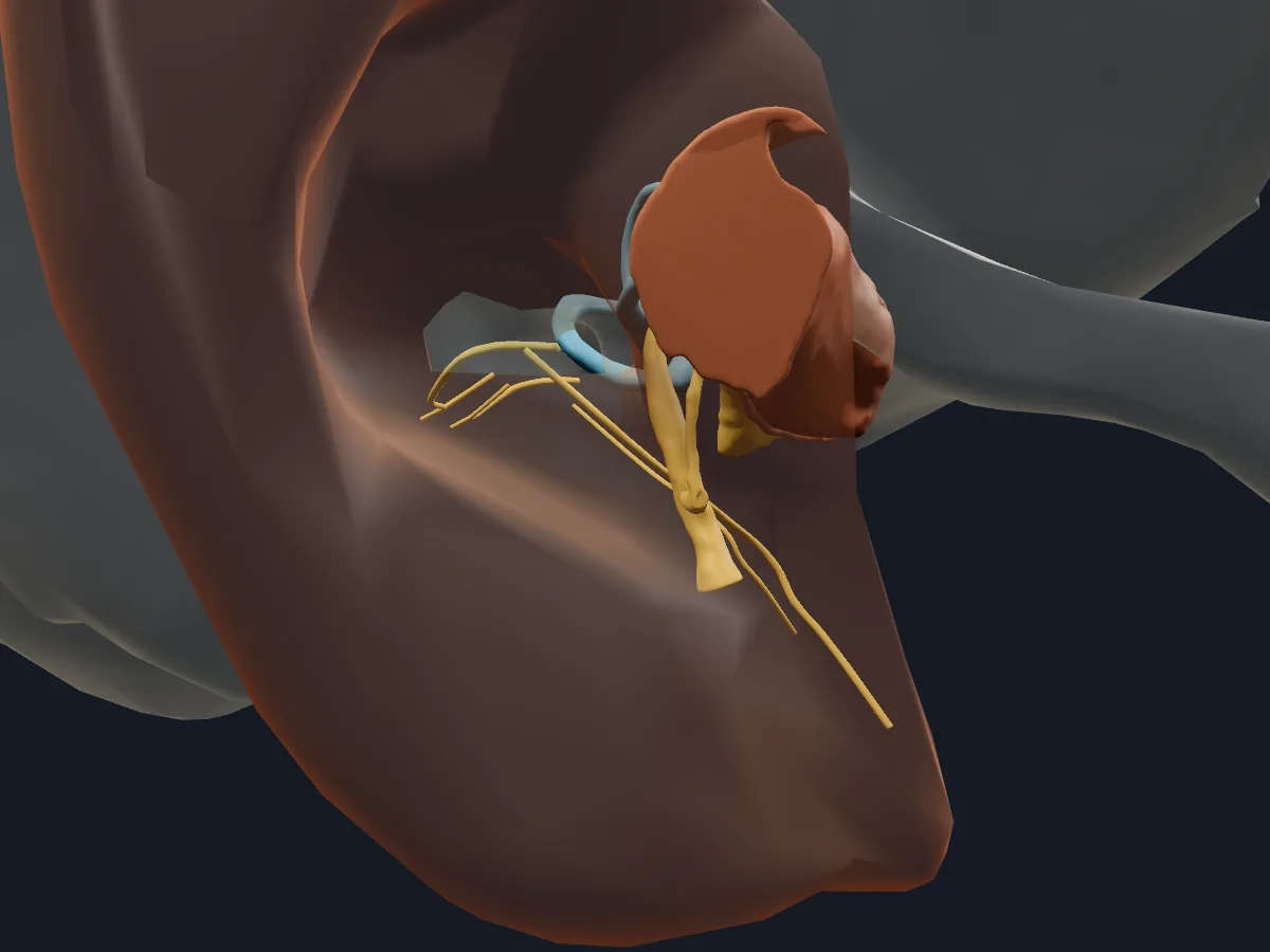
The middle and inner ear in place inside the temporal bone, with the external acoustic meatus.
Open this view in 3DImage: Cogni Anatomy · CC BY-SA 4.0 · Sources: BodyParts3D, OpenEar, Z-Anatomy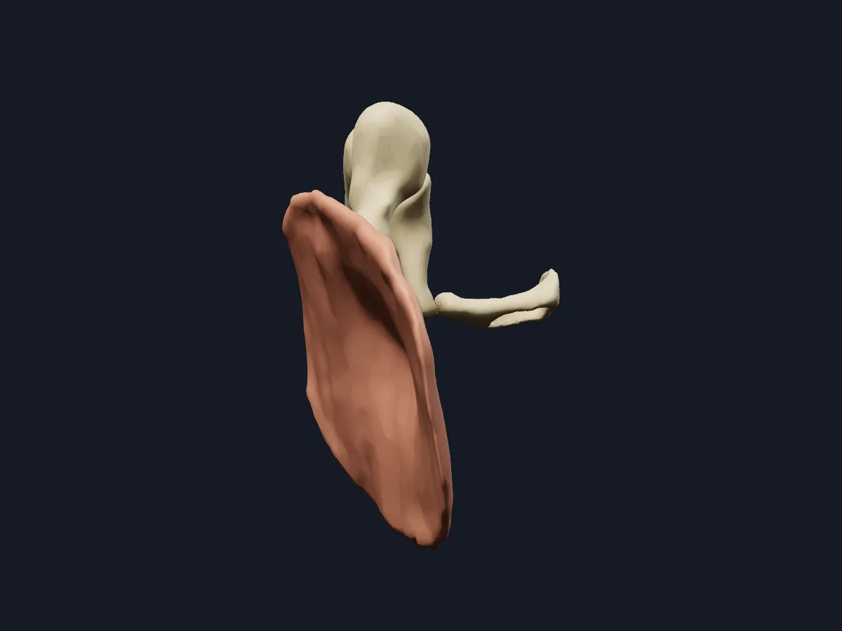
The malleus, incus and stapes enlarged to show their shape and how they connect.
Open this view in 3DImage: Cogni Anatomy · CC BY 4.0 · Sources: OpenEar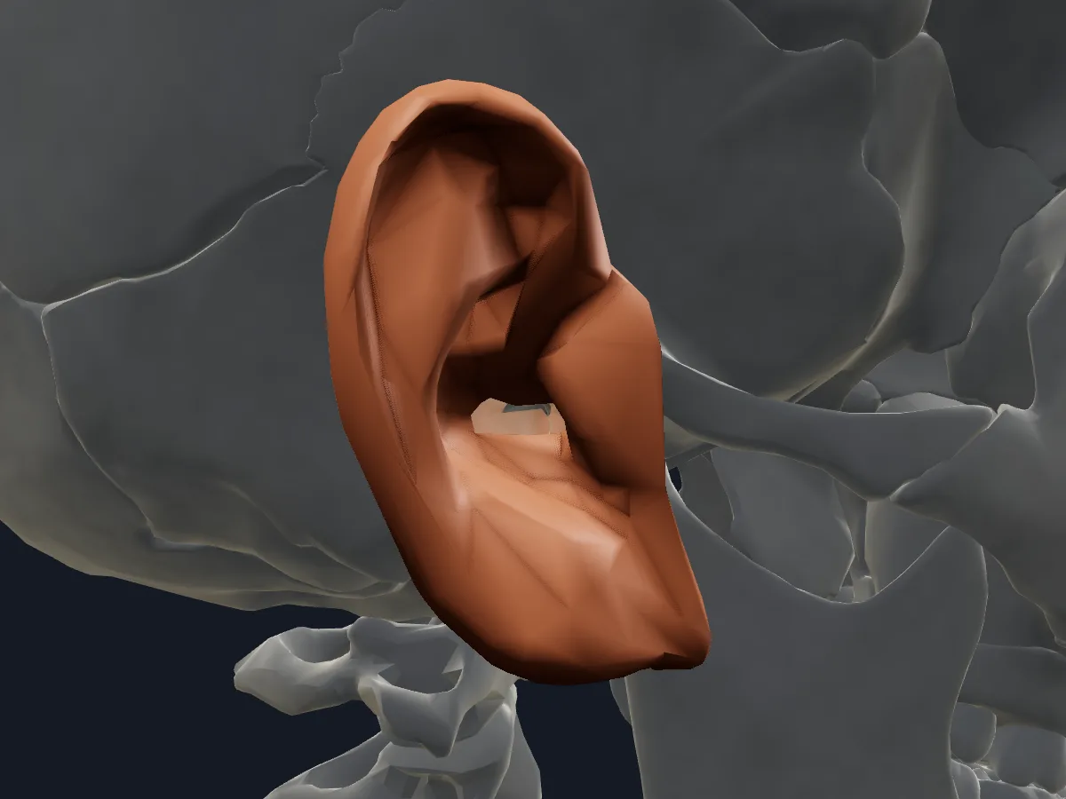
The auricle of the external ear on its own, with the skull faded behind it.
Open this view in 3DImage: Cogni Anatomy · CC BY 4.0 · Sources: BodyParts3D
What is in this model
15 3D parts. Names follow the Foundational Model of Anatomy (FMA).
- Nerves 7
- Sense organs 5
- Bones 3
- Chorda tympani
- External acoustic meatus
- External ear
- Facial nerve (temporal part)
- Incus
- Malleus
- Scala tympani (cochlea)
- Scala vestibuli and vestibule
- Stapes
- Tympanic membrane
- Vestibulocochlear nerve
- Vestibulocochlear nerve (VIII)
Questions
What are the three parts of the ear?
The external ear (auricle and external acoustic meatus), the middle ear (tympanic cavity with the ossicles) and the inner ear (vestibule, semicircular canals and cochlea).
What is the smallest bone in the human body?
The stapes in the middle ear, about 3 mm long. Its footplate fits into the oval window of the inner ear.
What do the ear ossicles do?
The malleus, incus and stapes form a chain that carries vibrations of the tympanic membrane to the oval window and amplifies their pressure for the fluid of the inner ear.