Eye anatomy in 3D
The eye is the organ of vision: an eyeball in the bony orbit with a fibrous outer coat, a vascular middle coat and the retina inside, enclosing the lens and vitreous body. It is moved by six extraocular muscles, connected to the brain by the optic nerve and kept moist by the lacrimal apparatus. In this free 3D model you can see the eyeball with its muscles, nerves and tear ducts in the orbit, cut through it and isolate each part.
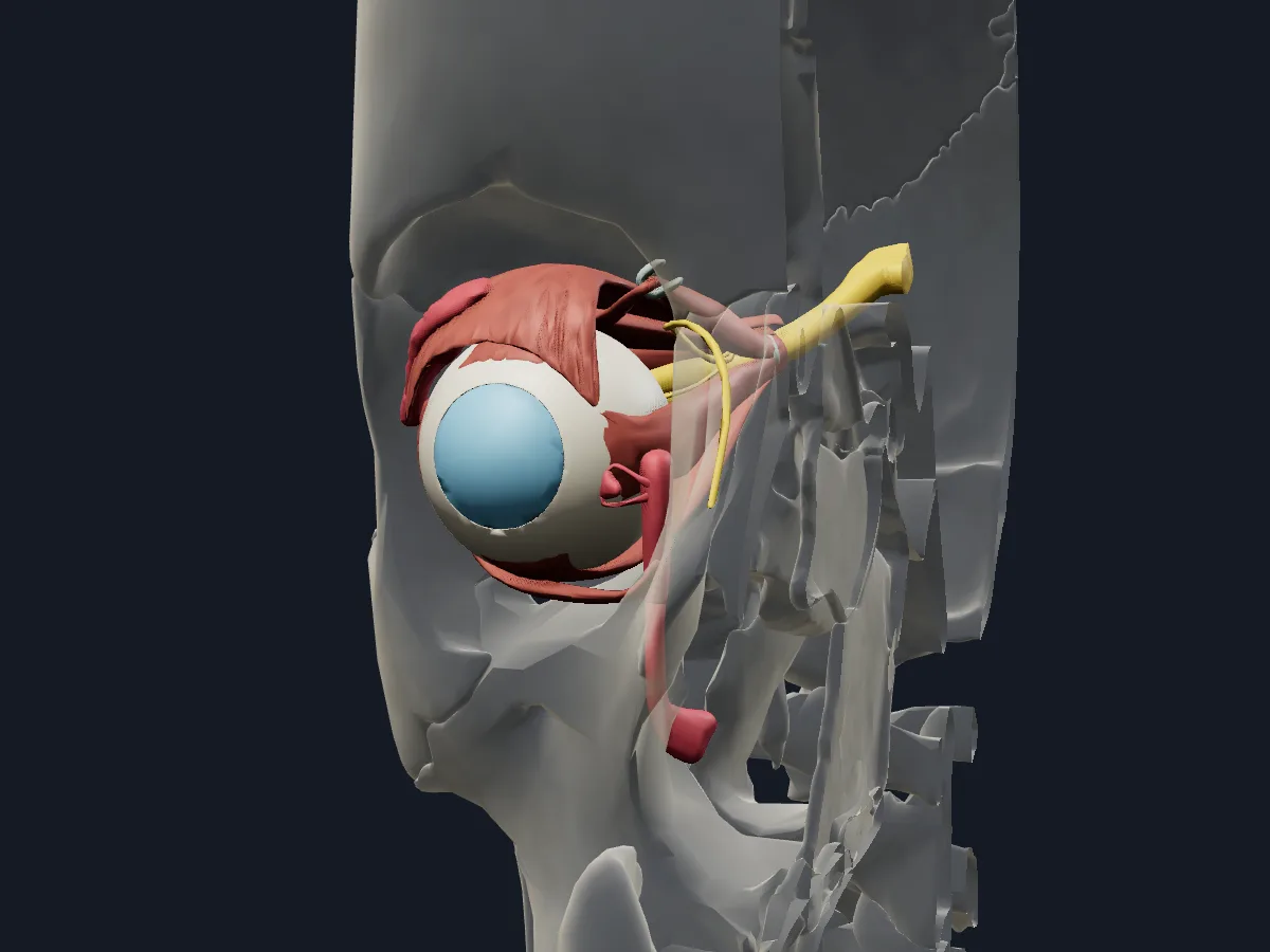 Image: Cogni Anatomy · CC BY 4.0 · Sources: BodyParts3D
- The adult eyeball is about 24 mm in diameter
- 3 coats: fibrous (sclera and cornea), vascular (choroid, ciliary body and iris) and inner (retina)
- 6 extraocular muscles: 4 recti and 2 obliques
- The 4 recti arise from the common tendinous ring at the apex of the orbit
- The nasolacrimal duct carries tears into the inferior nasal meatus
Structure of the eye
Light passes through the cornea, the aqueous humour of the anterior chamber, the pupil in the iris, the lens and the vitreous body before reaching the retina. The lens hangs from the ciliary body by the suspensory ligament, and the ciliary muscle changes its shape for near and far vision. The optic nerve leaves the back of the eyeball slightly medial to the posterior pole.
The superior, inferior, medial and lateral recti and the superior and inferior obliques move the eyeball, and the levator palpebrae superioris raises the upper eyelid. Tears from the lacrimal gland drain through the lacrimal canaliculi into the lacrimal sac and nasolacrimal duct.
What the 3D model shows
The view shows the right eye in its orbit with the skull faded around it, 32 parts in total. The eyeball is built from the sclera, cornea, choroid, iris, ciliary body, anterior chamber, lens with its suspensory ligament, vitreous body and retina. Around it are the four recti, the two obliques with the trochlea of the superior oblique, the levator palpebrae superioris and the common tendinous ring. The optic nerve, ciliary ganglion, nasociliary nerve and long and short ciliary nerves are included, along with the lacrimal gland, lacrimal lake, canaliculus, lacrimal sac and nasolacrimal duct.
How to study the eye
From the superior view, follow the four recti back to the common tendinous ring and the superior oblique forward to its trochlea, where it turns back to the eyeball. Use the section tool with a horizontal plane through the eyeball and name the structures from the cornea to the retina. Hide the sclera to see the choroid underneath. In the anterior view, trace the tear path from the lacrimal gland to the nasolacrimal duct. Then test yourself on each extraocular muscle: its action and its nerve.
What is in this model
32 3D parts. Names follow the Foundational Model of Anatomy (FMA).
- Sense organs 16
- Nerves 7
- Muscles 7
- Connective tissue 2
- Anterior chamber of right eyeball
- Choroid
- Ciliary ganglion
- Common tendinous ring
- Communicating branch of right nasociliary nerve with right ciliary ganglion
- Cornea
- Corona ciliaris
- Inferior oblique
- Inferior rectus
- Iris
- Lacrimal canaliculus
- Lacrimal gland
- Lacrimal lake
- Lacrimal sac
- Lateral rectus
- Lens
- Levator palpebrae superioris
- Long ciliary nerve
- Medial rectus
- Nasociliary nerve
- Nasolacrimal duct
- Optic nerve
- Optic part of right retina
- Sclera
- Short ciliary nerve
- Superior oblique
- Superior rectus
- Suspensory ligament of right lens
- Trochlea of right superior oblique
- Vitreous body
Questions
What are the layers of the eye?
Three coats: the outer fibrous coat (sclera and cornea), the middle vascular coat (choroid, ciliary body and iris) and the inner coat, the retina.
How many muscles move the eye?
Six: the superior, inferior, medial and lateral recti and the superior and inferior obliques. The levator palpebrae superioris, also in the orbit, raises the upper eyelid rather than moving the eyeball.
Where do tears drain?
From the lacrimal lake at the medial angle of the eye through the lacrimal canaliculi into the lacrimal sac, then through the nasolacrimal duct into the inferior nasal meatus.