Skull anatomy in 3D
The skull is the skeleton of the head: a cranium that encloses the brain and a facial skeleton that frames the orbits, nasal cavity and mouth. Its bones are joined by immovable sutures, except the mandible, which moves at the temporomandibular joint. In this free 3D model you can turn the skull in any direction, give each bone its own colour and lift out the mandible to study it alone.
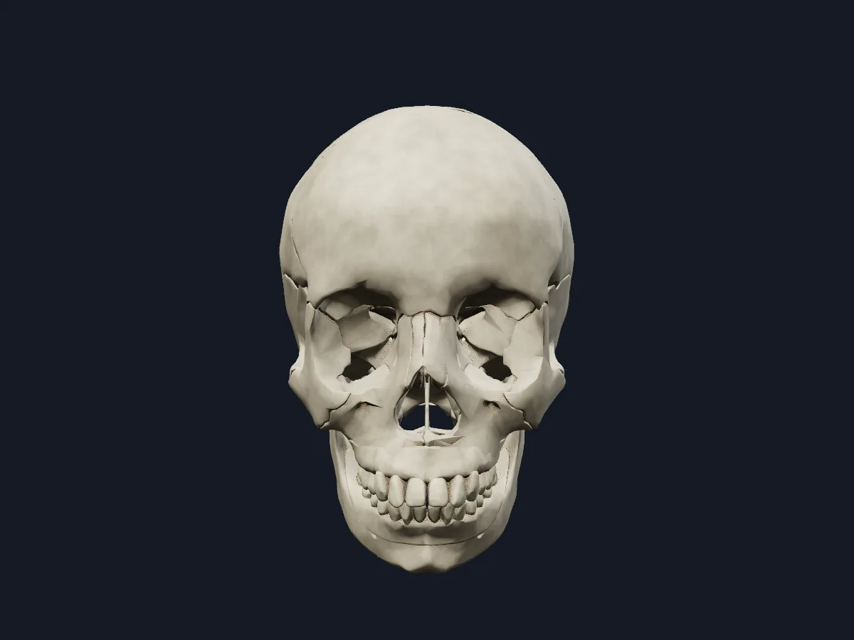 Image: Cogni Anatomy · CC BY 4.0 · Sources: BodyParts3D
- 22 bones: 8 cranial and 14 facial
- The mandible is the only skull bone that moves, at the temporomandibular joint
- Main sutures of the vault: coronal, sagittal, lambdoid and squamous
- The cranial base has three steps: the anterior, middle and posterior cranial fossae
- The foramen magnum in the occipital bone is where the brainstem continues into the spinal cord
Cranial and facial bones
The cranium (neurocranium) is built from the frontal, ethmoid, sphenoid and occipital bones, which are unpaired, and the paired parietal and temporal bones. The flat bones form the vault, while the sphenoid, ethmoid, temporal and occipital bones form the cranial base.
The facial skeleton (viscerocranium) includes the paired maxillae, zygomatic, nasal, lacrimal and palatine bones and inferior nasal conchae, and the unpaired vomer and mandible. The maxillae carry the upper teeth and form most of the hard palate; the mandible carries the lower teeth.
What the 3D model shows
The skull view shows the frontal, parietal, occipital, temporal, sphenoid and ethmoid bones, the maxillae, mandible, nasal, zygomatic and palatine bones and the vomer, together with the upper and lower permanent teeth from the central incisors to the second molars, the gums and the alar cartilages of the nose. A second view colours the bones so their borders stand out. The head view adds the cervical vertebrae with their intervertebral discs, the hyoid bone and the cartilages of the larynx, and a separate view isolates the mandible.
How to study the skull
Open the coloured view and start from the front: name the bones around the orbit and the nasal opening. Turn to the lateral view to find where the frontal, parietal, temporal and sphenoid bones meet at the pterion, then look from above to follow the coronal, sagittal and lambdoid sutures. From below, find the foramen magnum, the palatine bones and the vomer. Use the explode slider to separate the bones, then isolate the mandible and name its body, ramus and processes. Close the model and list all 22 bones from memory.
Views in the 3D model
The skull with its cranial and facial bones, teeth and gums.
Open this view in 3DImage: Cogni Anatomy · CC BY 4.0 · Sources: BodyParts3D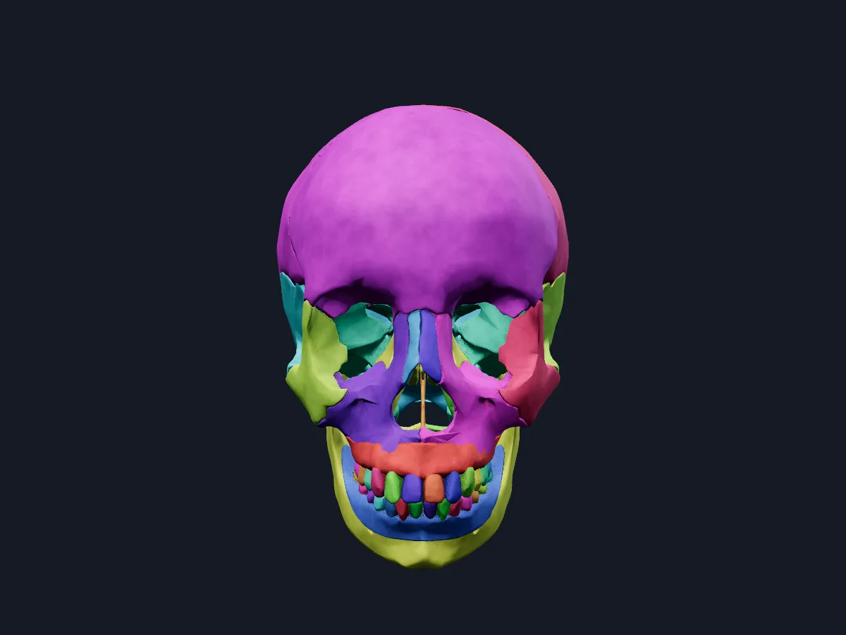
The same skull with the bones coloured so each one and its sutures stand out.
Open this view in 3DImage: Cogni Anatomy · CC BY 4.0 · Sources: BodyParts3D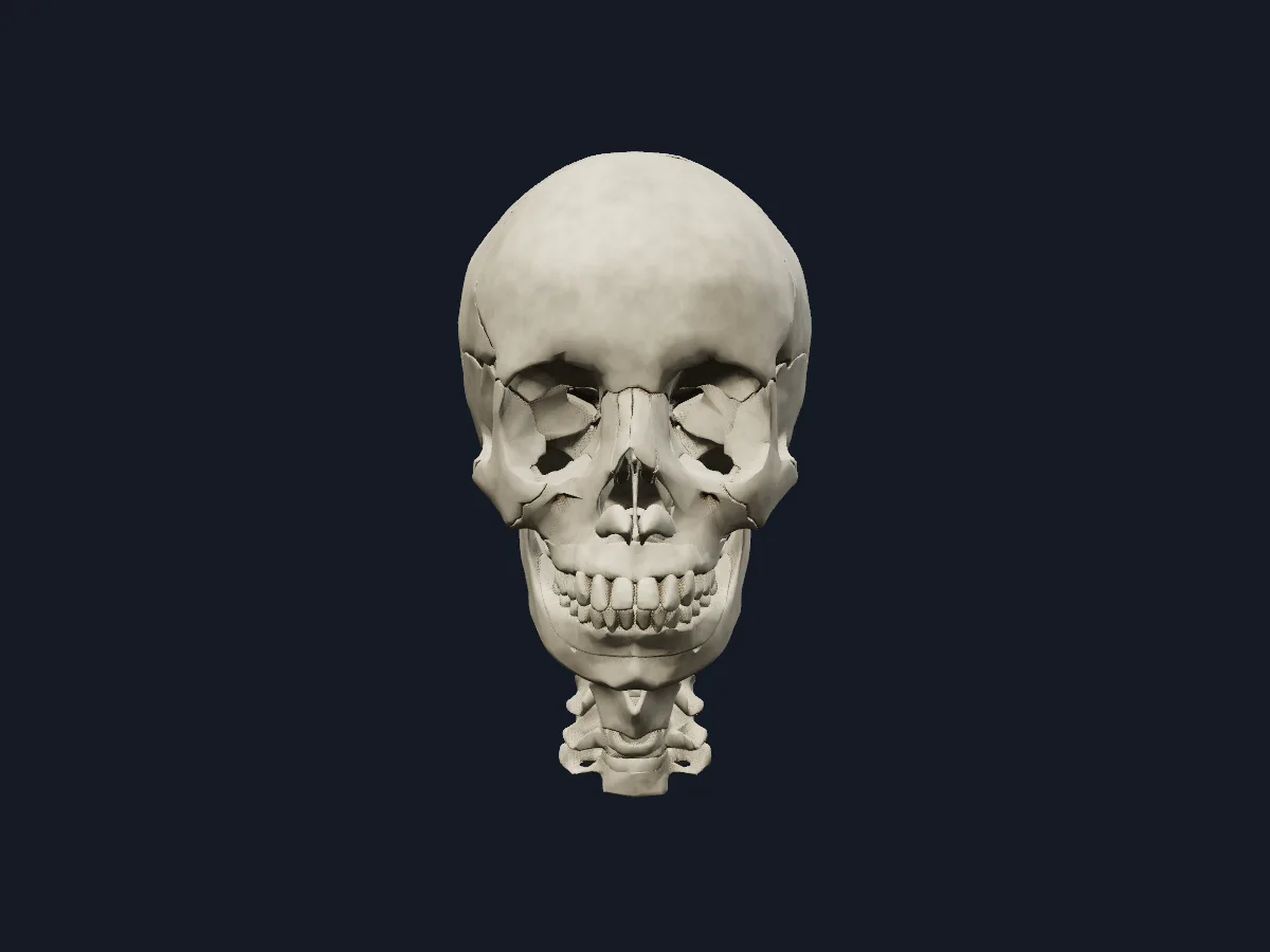
The skull together with the cervical vertebrae, intervertebral discs, hyoid bone and laryngeal cartilages.
Open this view in 3DImage: Cogni Anatomy · CC BY 4.0 · Sources: BodyParts3D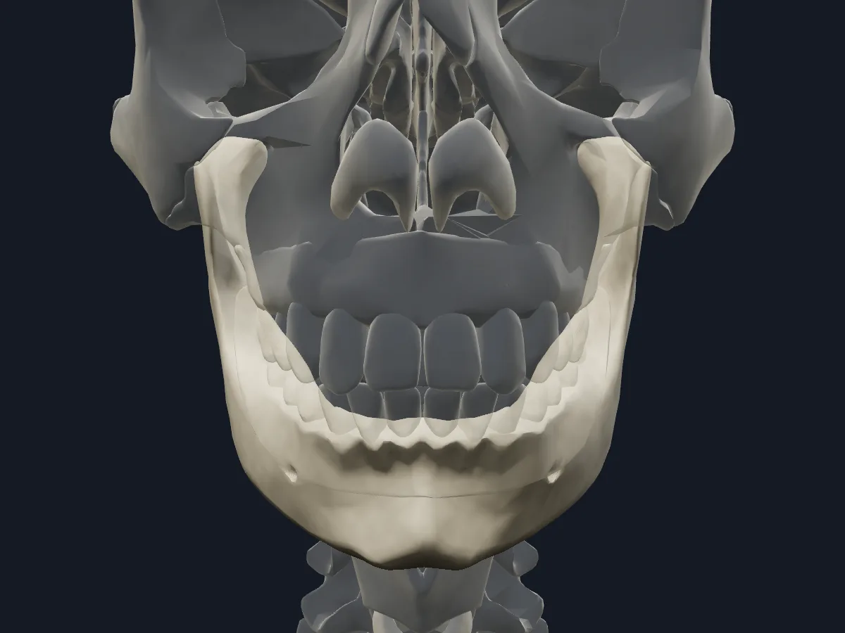
The mandible on its own, with the rest of the skeleton faded for orientation.
Open this view in 3DImage: Cogni Anatomy · CC BY 4.0 · Sources: BodyParts3D
What is in this model
73 3D parts. Names follow the Foundational Model of Anatomy (FMA).
- Bones 73
- Arytenoid cartilage
- Atlas
- Axis
- Corniculate cartilage
- Cricoid cartilage
- Cuneiform cartilage
- Ethmoid
- Fifth cervical vertebra
- Fourth cervical vertebra
- Frontal bone
- Gingiva of lower jaw
- Gingiva of upper jaw
- Hyoid bone
- Intervertebral disk of axis
- Intervertebral disk of fifth cervical vertebra
- Intervertebral disk of fourth cervical vertebra
- Intervertebral disk of sixth cervical vertebra
- Intervertebral disk of third cervical vertebra
- Lower central secondary incisor tooth
- Lower first secondary molar tooth
- Lower first secondary premolar tooth
- Lower lateral secondary incisor tooth
- Lower second secondary molar tooth
- Lower second secondary premolar tooth
- Lower secondary canine tooth
- Major alar cartilage
- Mandible
- Maxilla
- Nasal bone
- Occipital bone
- Palatine bone
- Parietal bone
- Seventh cervical vertebra
- Sixth cervical vertebra
- Sphenoid bone
- Temporal bone
- Third cervical vertebra
- Thyroid cartilage
- Upper central secondary incisor tooth
- Upper first secondary molar tooth
- and 7 more
Questions
How many bones are in the human skull?
22: 8 cranial bones and 14 facial bones. Counting the six auditory ossicles inside the temporal bones gives 28; the hyoid bone is usually described separately.
Which bone of the skull can move?
Only the mandible. It articulates with the temporal bones at the two temporomandibular joints, which open and close the mouth and allow chewing.
Which bones form the cranial vault?
Mainly the frontal bone, the two parietal bones, the occipital bone and the squamous parts of the temporal bones, with the greater wings of the sphenoid at the sides.