Facial muscles in 3D
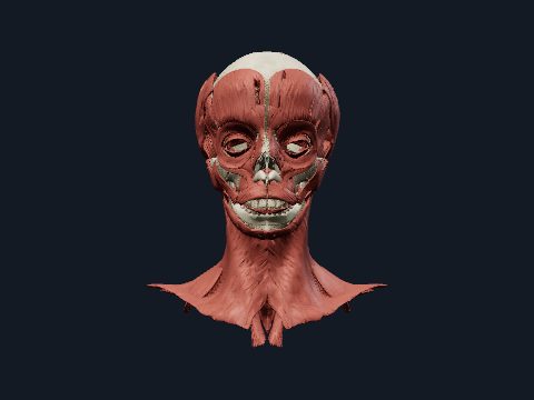
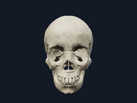
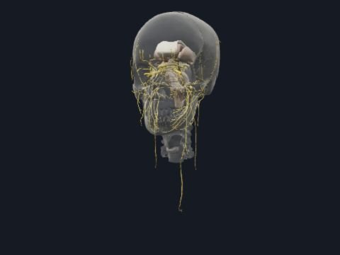
The muscles of facial expression are thin muscles of the face and scalp that move the skin around the eyes, nose and mouth and are supplied by the facial nerve. The muscles of mastication are a separate, deeper group that move the mandible in chewing and are supplied by the mandibular nerve. This free 3D model shows both groups over a see-through skull, so you can isolate each muscle and see which bone it arises from.
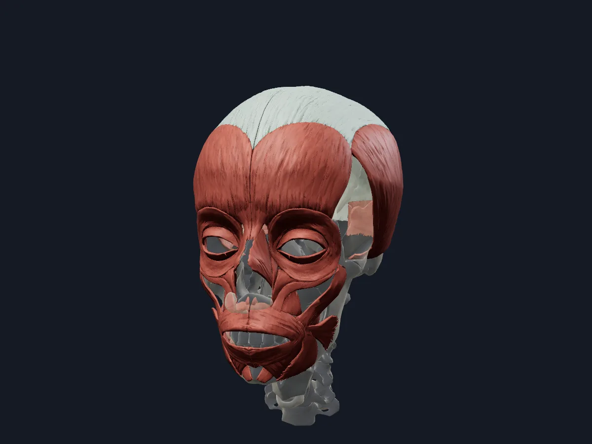 Image: Cogni Anatomy · CC BY-SA 4.0 · Sources: BodyParts3D, Z-Anatomy
- Muscles of facial expression are supplied by the facial nerve (CN VII)
- They develop from the second pharyngeal arch
- 4 muscles of mastication: masseter, temporalis, medial and lateral pterygoid
- Muscles of mastication are supplied by the mandibular nerve (V3) and develop from the first pharyngeal arch
- The buccinator forms the muscular wall of the cheek and is a facial muscle, not a muscle of mastication
Muscles of expression and mastication
Most muscles of facial expression arise from bone or fascia and insert into the skin, grouped around the openings of the face. The orbicularis oculi closes the eyelids and the orbicularis oris closes the lips; muscles such as the zygomaticus major and minor, levator labii superioris, levator anguli oris, risorius, depressor anguli oris and depressor labii inferioris pull the lips in different directions. The frontalis and occipitalis are joined by the epicranial aponeurosis.
Of the masticatory muscles, the temporalis, masseter and medial pterygoid close the jaw, while the lateral pterygoid pulls the mandible forward and helps open the mouth.
What the 3D model shows
The facial muscles view shows both sides of the face and scalp: the frontalis, occipitalis and temporoparietalis with the epicranial aponeurosis, the orbital and palpebral parts of the orbicularis oculi, the corrugator supercilii and procerus, the nasalis and depressor septi nasi, the orbicularis oris, buccinator, mentalis and the elevators and depressors of the lips and mouth angle. The mastication view shows the temporalis, the superficial and deep parts of the masseter, the medial pterygoid and the superior and inferior heads of the lateral pterygoid on both sides. In both views the skull is faded for orientation.
How to study the facial muscles
In the facial muscles view, start from the anterior view with the two sphincters, orbicularis oculi and orbicularis oris, then add the muscles that radiate from the mouth one at a time. Turn to the lateral view to see the buccinator deep to the cheek muscles. In the mastication view, hide the masseter and temporalis to reach the two pterygoids on the inner side of the mandibular ramus. For each muscle, say its action, then test yourself on which nerve supplies it, facial or mandibular.
Views in the 3D model
Muscles of facial expression on the face and scalp, with the skull faded in the background.
Open this view in 3DImage: Cogni Anatomy · CC BY-SA 4.0 · Sources: BodyParts3D, Z-Anatomy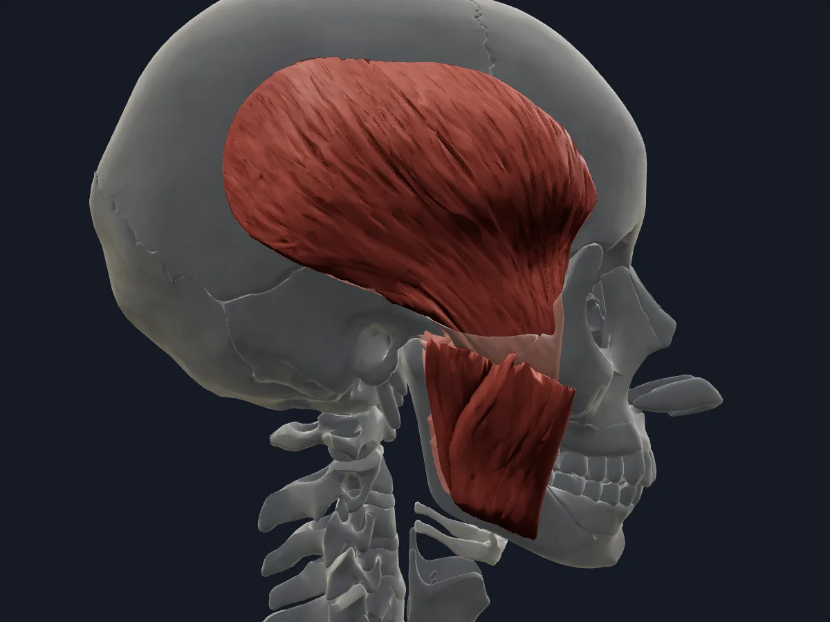
The temporalis, masseter and pterygoid muscles that move the mandible, over a faded skull.
Open this view in 3DImage: Cogni Anatomy · CC BY-SA 4.0 · Sources: BodyParts3D, Z-Anatomy
What is in this model
54 3D parts. Names follow the Foundational Model of Anatomy (FMA).
- Muscles 52
- Connective tissue 2
- Buccinator
- Corrugator supercilii
- Deep part of masseter
- Depressor anguli oris
- Depressor labii inferioris
- Depressor septi nasi
- Epicranial aponeurosis
- Frontalis muscle
- Inferior head of lateral pterygoid muscle
- Levator anguli oris
- Levator labii superioris
- Levator nasolabialis
- Medial pterygoid muscle
- Mentalis muscle
- Nasalis muscle
- Occipitalis muscle
- Orbicularis oris muscle
- Orbital part of orbicularis oculi
- Palpebral part of orbicularis oculi
- Procerus muscle
- Risorius muscle
- Superficial part of masseter
- Superior head of lateral pterygoid muscle
- Temporalis muscle
- Temporoparietalis muscle
- Zygomaticus major muscle
- Zygomaticus minor muscle
Questions
Which nerve supplies the muscles of facial expression?
The facial nerve (CN VII). After leaving the skull it divides in the parotid gland into temporal, zygomatic, buccal, marginal mandibular and cervical branches.
What are the muscles of mastication?
The masseter, temporalis, medial pterygoid and lateral pterygoid. All four attach to the mandible and are supplied by the mandibular division of the trigeminal nerve.
Is the buccinator a muscle of mastication?
No. It is a muscle of facial expression supplied by the facial nerve, although it helps chewing by pressing the cheek against the teeth and keeping food between them.