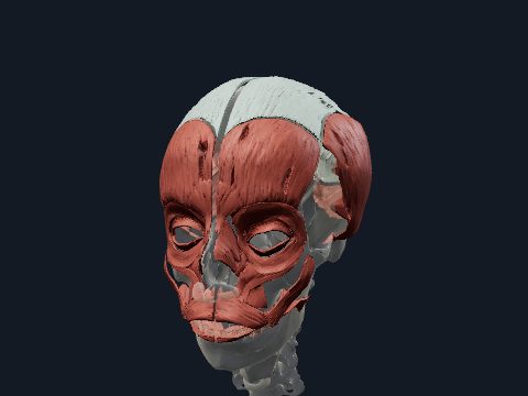
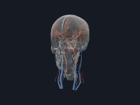
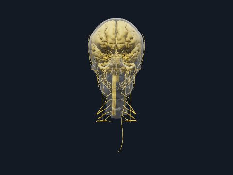
Head and neck muscles in 3D
The head and neck muscles move the face, jaw, tongue, pharynx and larynx and hold and turn the head on the cervical spine. They fall into facial and masticatory muscles, suprahyoid and infrahyoid groups, the sternocleidomastoid and scalenes, deep prevertebral muscles and the posterior neck muscles down to the suboccipital group. In this free 3D model you can view all of them over the skull and cervical vertebrae, or together with every other system of the head, and isolate any single muscle.
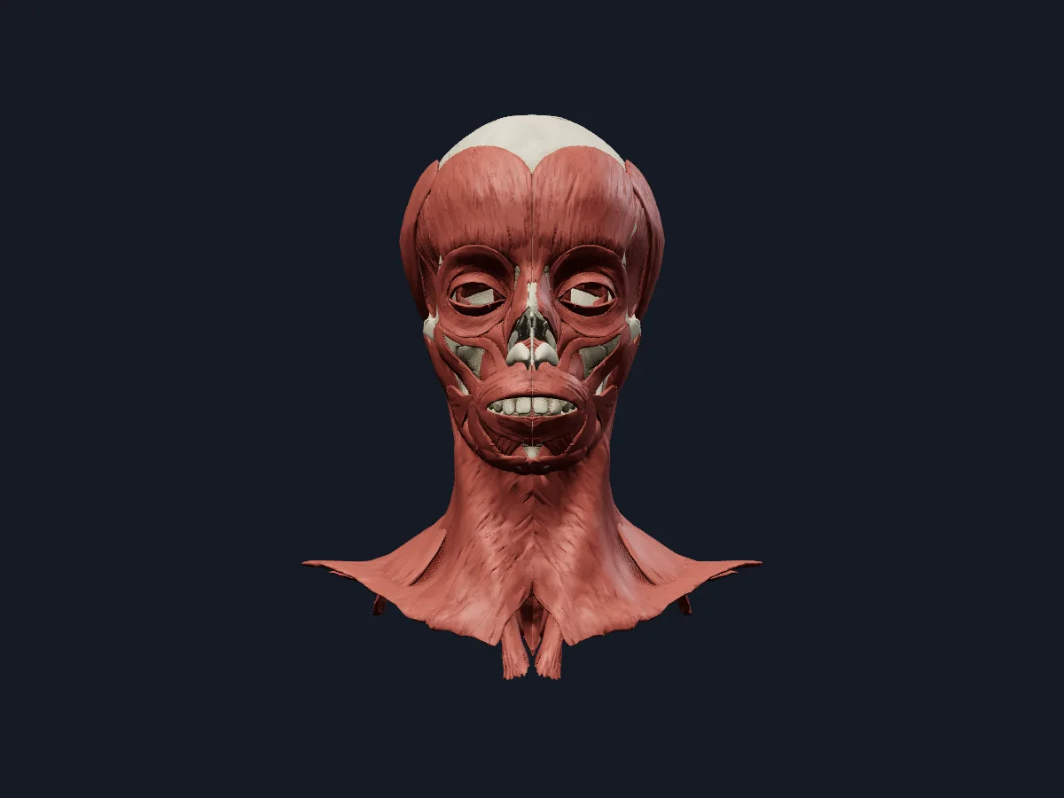 Image: Cogni Anatomy · CC BY-SA 4.0 · Sources: BodyParts3D, Z-Anatomy
- The sternocleidomastoid divides each side of the neck into anterior and posterior triangles
- 4 suprahyoid muscles: digastric, stylohyoid, mylohyoid and geniohyoid
- 4 infrahyoid muscles: sternohyoid, sternothyroid, thyrohyoid and omohyoid
- 3 scalene muscles: anterior, middle and posterior
- 4 suboccipital muscles: rectus capitis posterior major and minor, obliquus capitis superior and inferior
Muscle groups of the head and neck
Superficially lie the platysma, the sternocleidomastoid and the upper part of the trapezius. In front, the hyoid bone separates the suprahyoid muscles, which form the floor of the mouth and raise the hyoid, from the infrahyoid strap muscles, which lower it with the larynx. Laterally, the scalenes run from the cervical transverse processes to the first and second ribs; the longus colli and longus capitis lie directly on the vertebral bodies.
At the back, the splenius capitis, semispinalis capitis and longissimus capitis extend the head, and the small suboccipital muscles connect the atlas, axis and occipital bone. Inside lie the muscles of the tongue, soft palate, pharynx and larynx.
What the 3D model shows
The muscles and bones view places every muscle of the head and neck on the skull, cervical vertebrae, hyoid bone and laryngeal cartilages. It includes the platysma, sternocleidomastoid and descending part of the trapezius, the digastric with its intermediate tendon, the mylohyoid, geniohyoid, stylohyoid, omohyoid and thyrohyoid, the scalenes and prevertebral muscles, the suboccipital and deep posterior neck muscles, the pharyngeal constrictors, the tongue and soft palate muscles, the intrinsic laryngeal muscles including the vocalis, and the extraocular muscles. The all-systems view adds the arteries, veins, nerves, brain, eyes and glands of the head, about 750 parts in total.
How to study the head and neck muscles
Begin in the lateral view and find the sternocleidomastoid; everything in the neck is described in relation to it. Hide it to see the scalenes and the splenius capitis behind. In the anterior view, use the hyoid bone as the centre: name the muscles above it, then those below. From the posterior view, peel off the trapezius, splenius and semispinalis with the muscle layer control to reach the suboccipital muscles. Finally, switch to the all-systems view and follow the hypoglossal nerve towards the tongue. Then name each group from memory with its action.
Views in the 3D model
All muscles of the head and neck on the skull, cervical spine, hyoid bone and laryngeal cartilages.
Open this view in 3DImage: Cogni Anatomy · CC BY-SA 4.0 · Sources: BodyParts3D, Z-Anatomy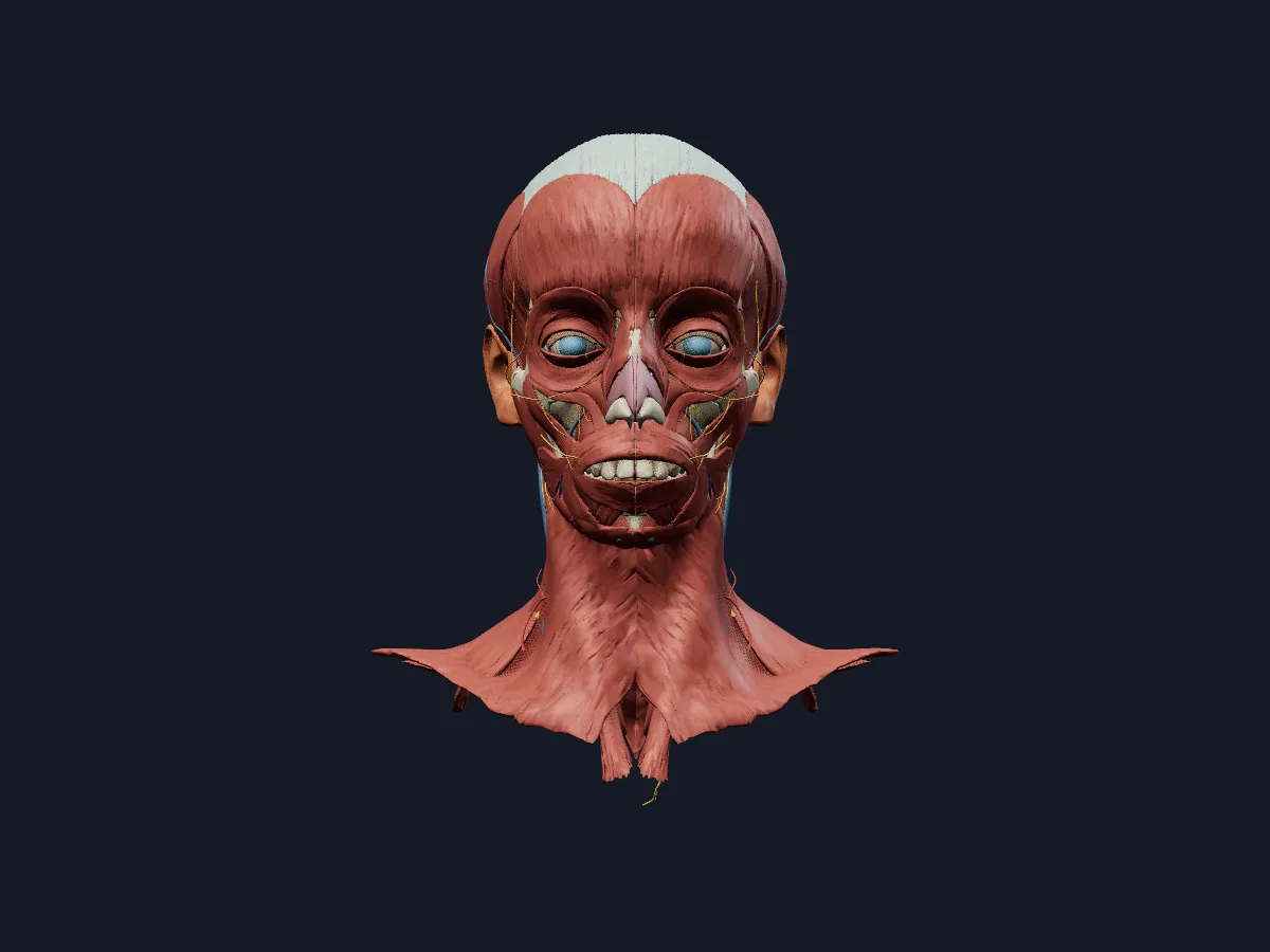
The head and neck with every system shown: bones, muscles, vessels, nerves, brain, eyes and glands.
Open this view in 3DImage: Cogni Anatomy · CC BY-SA 4.0 · Sources: BodyParts3D, HRA, Z-Anatomy
What is in this model
751 3D parts. Names follow the Foundational Model of Anatomy (FMA).
- Nerves 200
- Muscles 181
- Arteries 157
- Bones 73
- Veins 57
- Sense organs 39
- Connective tissue 31
- Respiratory 6
- Digestive 5
- Endocrine 2
- Abducens nerve (VI)
- Amygdala
- Angular gyrus
- Angular vein
- Anterior belly of digastric muscle
- Anterior cerebral artery
- Anterior chamber of left eyeball
- Anterior chamber of right eyeball
- Anterior choroidal artery
- Anterior commissure
- Anterior communicating artery
- Anterior division of mandibular nerve
- Anterior division of retromandibular vein
- Anterior ethmoidal nerve
- Anterior inferior cerebellar artery
- Anterior intercavernous sinus
- Anterior parietal artery
- Anterior part of left superior temporal gyrus
- Anterior part of right superior temporal gyrus
- Anterior spinal artery
- Anterior temporal artery
- Anterior temporal branch of left lateral occipital artery
- Anterior temporal branch of right lateral occipital artery
- Anterolateral central branch of left middle cerebral artery
- Anterolateral central branch of right middle cerebral artery
- Artery of central sulcus
- Artery of left postcentral sulcus
- Artery of left precentral sulcus
- Artery of right postcentral sulcus
- Artery of right precentral sulcus
- Ary-epiglottic part of oblique arytenoid muscle
- Aryepiglotticus
- Arytenoid cartilage
- Atlas
- Axis
- Basilar artery
- Basilar venous plexus
- Brachium of left inferior colliculus
- Brachium of left superior colliculus
- Brachium of right inferior colliculus
- and 389 more
Questions
Which muscle turns the head?
Mainly the sternocleidomastoid. Acting alone it tilts the head to its own side and turns the face towards the opposite side; acting together the two muscles flex the neck.
Which muscles move the hyoid bone?
The suprahyoid muscles (digastric, stylohyoid, mylohyoid, geniohyoid) raise it during swallowing, and the infrahyoid muscles (sternohyoid, sternothyroid, thyrohyoid, omohyoid) pull it and the larynx back down.
What are the suboccipital muscles?
Four small muscles on each side below the occipital bone: rectus capitis posterior major and minor and obliquus capitis superior and inferior. They extend and rotate the head at the atlanto-occipital and atlanto-axial joints.