Head and neck blood vessels in 3D
The brain is supplied by the two internal carotid arteries and the two vertebral arteries, which are linked at the base of the brain by the cerebral arterial circle (circle of Willis). Blood from the brain drains through the dural venous sinuses into the internal jugular veins, while the face and scalp drain through superficial veins such as the facial and retromandibular veins. In this free 3D model you can view arteries and veins together or separately inside a see-through skull and find any vessel by name.
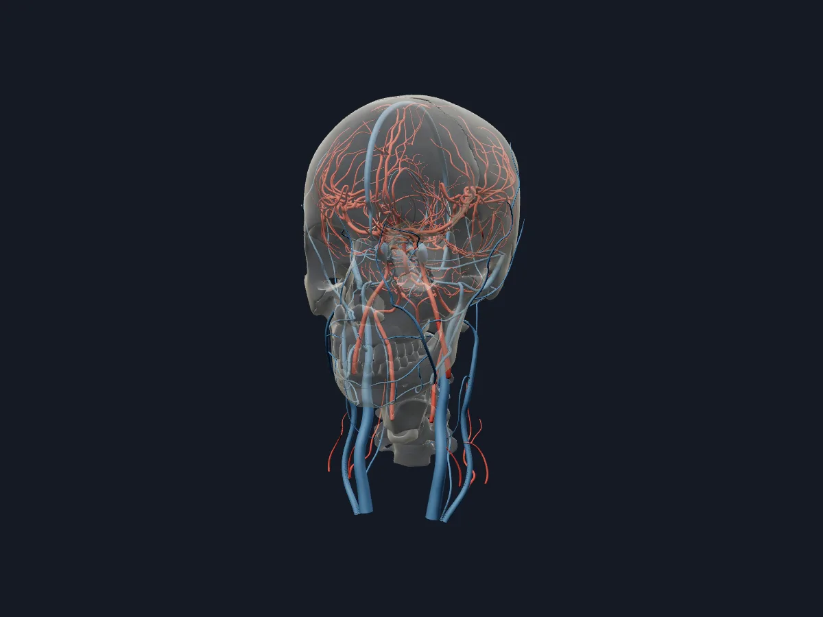 Image: Cogni Anatomy · CC BY-SA 4.0 · Sources: BodyParts3D, Z-Anatomy
- 4 arteries supply the brain: the paired internal carotid and vertebral arteries
- The two vertebral arteries unite at the lower border of the pons to form the basilar artery
- The circle of Willis is formed by the anterior cerebral, anterior communicating, internal carotid, posterior communicating and posterior cerebral arteries
- The sigmoid sinus continues as the internal jugular vein at the jugular foramen
- The superior sagittal sinus runs along the attached upper margin of the falx cerebri
Arteries and veins of the head
Each internal carotid artery gives off the ophthalmic artery and ends by dividing into the anterior and middle cerebral arteries. The anterior cerebral artery supplies the medial surface of the hemisphere, the middle cerebral artery most of the lateral surface. The vertebral arteries give the posterior inferior cerebellar arteries and join as the basilar artery, which gives the anterior inferior cerebellar, pontine and superior cerebellar arteries and ends as the two posterior cerebral arteries.
Venous blood collects in dural sinuses: the superior sagittal, straight and transverse sinuses lead to the sigmoid sinus and the internal jugular vein, and the cavernous sinus receives the ophthalmic veins.
What the 3D model shows
The arteries view has 157 parts: the internal carotid and vertebral arteries, the basilar artery, the complete circle of Willis, the middle cerebral artery with its insular part and cortical branches, the anterior cerebral artery with the pericallosal and callosomarginal arteries, the posterior cerebral, cerebellar, pontine and choroidal arteries, the ophthalmic and anterior spinal arteries, and the inferior thyroid, deep and superficial cervical arteries in the neck. The veins view has 57 parts: the dural venous sinuses, the internal and external jugular and vertebral veins, and the facial, angular, retromandibular, ophthalmic, lingual and other veins of the face and neck.
How to study the vessels of the head
Start with the arteries view from below. Follow each internal carotid artery up into the skull, then find the vertebral arteries joining into the basilar artery and the posterior communicating arteries closing the circle of Willis. Turn to the lateral view to follow the middle cerebral artery branches over the hemisphere, and to the medial side for the pericallosal artery. Then switch to the veins view, start at the superior sagittal sinus and trace the path to the internal jugular vein. Draw the circle of Willis from memory afterwards.
Views in the 3D model
Arteries and veins of the head and neck together inside a faded skull.
Open this view in 3DImage: Cogni Anatomy · CC BY-SA 4.0 · Sources: BodyParts3D, Z-Anatomy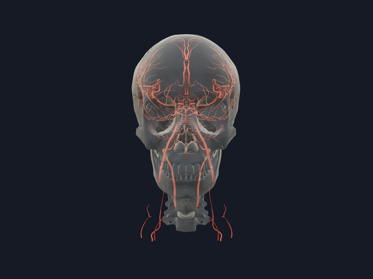
The arteries alone, from the internal carotid and vertebral arteries to the cortical branches of the cerebral arteries.
Open this view in 3DImage: Cogni Anatomy · CC BY 4.0 · Sources: BodyParts3D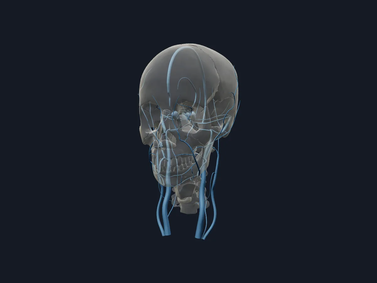
The veins alone: dural venous sinuses, jugular veins and the superficial veins of the face and neck.
Open this view in 3DImage: Cogni Anatomy · CC BY-SA 4.0 · Sources: BodyParts3D, Z-Anatomy
What is in this model
214 3D parts. Names follow the Foundational Model of Anatomy (FMA).
- Arteries 157
- Veins 57
- Angular vein
- Anterior cerebral artery
- Anterior choroidal artery
- Anterior communicating artery
- Anterior division of retromandibular vein
- Anterior inferior cerebellar artery
- Anterior intercavernous sinus
- Anterior parietal artery
- Anterior spinal artery
- Anterior temporal artery
- Anterior temporal branch of left lateral occipital artery
- Anterior temporal branch of right lateral occipital artery
- Anterolateral central branch of left middle cerebral artery
- Anterolateral central branch of right middle cerebral artery
- Artery of central sulcus
- Artery of left postcentral sulcus
- Artery of left precentral sulcus
- Artery of right postcentral sulcus
- Artery of right precentral sulcus
- Basilar artery
- Basilar venous plexus
- Branch of left anterior choroidal artery to posterior limb of left internal capsule
- Branch of left middle cerebral artery to left angular gyrus
- Branch of right anterior choroidal artery to posterior limb of right internal capsule
- Branch of right middle cerebral artery to right angular gyrus
- Callosomarginal artery
- Cavernous sinus
- Common facial vein
- Deep cervical artery
- External jugular vein
- Facial vein
- Hypothalamic branch of left posterior communicating artery
- Hypothalamic branch of right posterior communicating artery
- Inferior labial veins
- Inferior ophthalmic vein
- Inferior petrosal sinus
- Inferior sagittal sinus
- Inferior thyroid artery
- Insular part of left middle cerebral artery
- Insular part of right middle cerebral artery
- and 76 more
Questions
What is the circle of Willis?
An arterial ring at the base of the brain that links the internal carotid and vertebrobasilar systems. It is formed by the anterior and posterior cerebral arteries, the anterior and posterior communicating arteries and the ends of the internal carotid arteries.
Which arteries supply the brain?
The two internal carotid arteries supply most of the cerebrum, and the two vertebral arteries, through the basilar artery, supply the brainstem, cerebellum and occipital lobes.
Where do the dural venous sinuses drain?
Almost all of them drain, directly or indirectly, into the sigmoid sinuses, which leave the skull through the jugular foramina as the internal jugular veins.