Head and neck nerves in 3D
The head and neck are innervated mainly by the cranial nerves, which attach to the central nervous system and leave the skull through its foramina, and by the cervical spinal nerves. The trigeminal nerve carries sensation from the face, the facial nerve moves the face, and other cranial nerves serve the eye, ear, tongue, pharynx and larynx. In this free 3D model the nerves are shown with the brain and cervical spinal cord inside a see-through skull, and you can add the muscles and bones they run between.
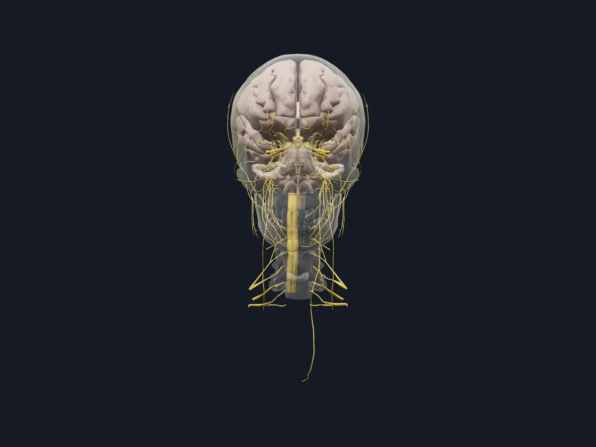 Image: Cogni Anatomy · CC BY-SA 4.0 · Sources: BodyParts3D, HRA, Z-Anatomy
- 12 pairs of cranial nerves, numbered I to XII
- The trigeminal nerve has 3 divisions: ophthalmic (V1), maxillary (V2) and mandibular (V3)
- 8 pairs of cervical spinal nerves, although there are only 7 cervical vertebrae
- The brachial plexus is formed by the anterior rami of C5–T1
- The chorda tympani, a branch of the facial nerve, joins the lingual nerve and carries taste from the anterior two-thirds of the tongue
How the head and neck are innervated
The olfactory, optic and vestibulocochlear nerves serve smell, vision, hearing and balance. The oculomotor, trochlear and abducens nerves move the eye. The trigeminal nerve supplies the skin of the face, the teeth and the mucosa of the nose and mouth through its three divisions, and its mandibular division also supplies the muscles of mastication. The facial nerve supplies the muscles of facial expression, the glossopharyngeal and vagus nerves the pharynx and larynx, and the hypoglossal nerve the muscles of the tongue.
Below the skull, the cervical spinal nerves supply the skin and muscles of the neck, and the roots of the brachial plexus pass towards the upper limb.
What the 3D model shows
The nerves view has 200 parts: the brain, brainstem and cerebellum, the spinal cord segments C1–C8 and T1, and the cranial nerves with their branches. The ophthalmic nerve is shown with the frontal, supra-orbital, supratrochlear, lacrimal, nasociliary, ethmoidal and ciliary nerves and the ciliary ganglion; the mandibular nerve with its buccal, lingual, inferior alveolar, mental and mylohyoid branches; the facial nerve with the chorda tympani. The roots of the brachial plexus are included. The nerves and muscles view adds the facial, masticatory, extraocular, tongue, palate, pharyngeal and suboccipital muscles with the bones of the head and neck.
How to study the nerves of the head
In the nerves view, isolate the trigeminal nerve and follow its three divisions: the ophthalmic towards the orbit and forehead, the maxillary towards the upper jaw, and the mandibular towards the lower jaw. Find where the chorda tympani joins the lingual nerve and follow the inferior alveolar nerve to the mental nerve. Then switch to the nerves and muscles view and match the oculomotor, trochlear and abducens nerves with the eye muscles they supply. Finally, name each cranial nerve from its point of attachment to the central nervous system without looking at the labels.
Views in the 3D model
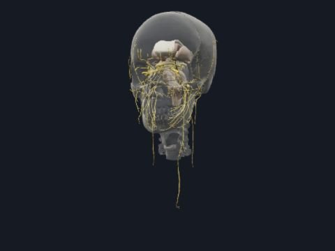
Cranial nerves and their branches with the brain and cervical spinal cord, inside a faded skull.
Open this view in 3DImage: Cogni Anatomy · CC BY-SA 4.0 · Sources: BodyParts3D, HRA, Z-Anatomy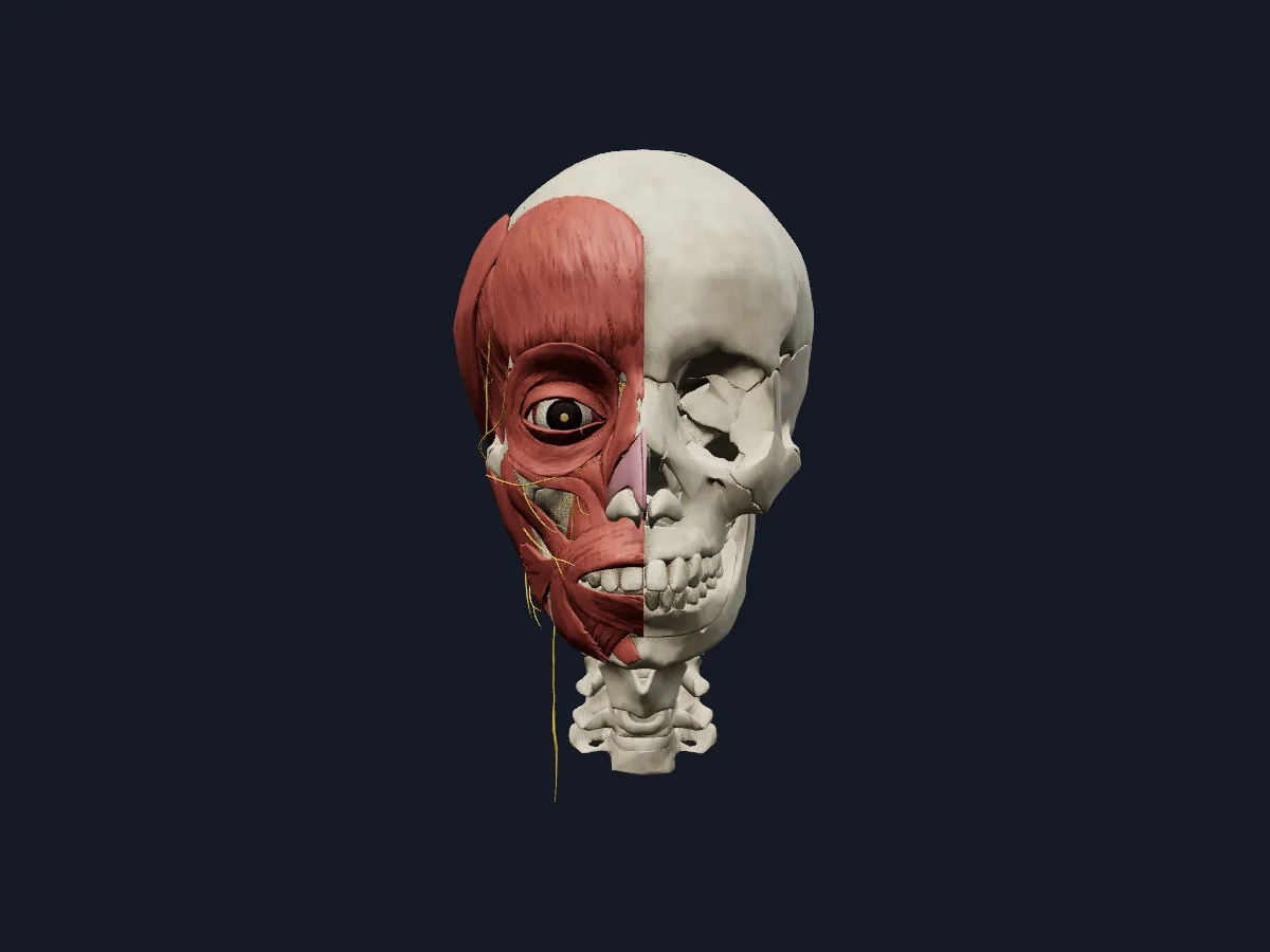
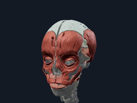
The same nerves shown with the muscles and bones of the head and neck.
Open this view in 3DImage: Cogni Anatomy · CC BY-SA 4.0 · Sources: BodyParts3D, HRA, Z-Anatomy
What is in this model
338 3D parts. Names follow the Foundational Model of Anatomy (FMA).
- Nerves 200
- Bones 73
- Muscles 58
- Sense organs 5
- Respiratory 2
- Abducens nerve (VI)
- Amygdala
- Angular gyrus
- Anterior belly of digastric muscle
- Anterior commissure
- Anterior division of mandibular nerve
- Anterior ethmoidal nerve
- Anterior part of left superior temporal gyrus
- Anterior part of right superior temporal gyrus
- Ary-epiglottic part of oblique arytenoid muscle
- Arytenoid cartilage
- Atlas
- Axis
- Brachium of left inferior colliculus
- Brachium of left superior colliculus
- Brachium of right inferior colliculus
- Brachium of right superior colliculus
- Buccal nerve
- Buccinator
- C1 segment of cervical spinal cord
- C2 segment of cervical spinal cord
- C3 segment of cervical spinal cord
- C4 segment of cervical spinal cord
- C5 segment of cervical spinal cord
- C6 segment of cervical spinal cord
- C7 segment of cervical spinal cord
- C8 segment of cervical spinal cord
- Caudate nucleus
- Central canal of spinal cord
- Cerebellum
- Cerebral aqueduct
- Chorda tympani
- Choroid
- Choroid plexus of cerebral hemisphere
- Ciliary ganglion
- Cingulate gyrus
- Commissure of fornix of forebrain
- Communicating branch of left nasociliary nerve with left ciliary ganglion
- Communicating branch of right nasociliary nerve with right ciliary ganglion
- Corniculate cartilage
- and 188 more
Questions
Which nerve supplies sensation to the face?
The trigeminal nerve (CN V). Its ophthalmic, maxillary and mandibular divisions supply the forehead and upper eyelid, the cheek and upper lip, and the lower lip and chin respectively.
What is the difference between the facial and the trigeminal nerve?
The facial nerve (CN VII) moves the muscles of facial expression, while the trigeminal nerve (CN V) carries sensation from the face and supplies the muscles of mastication.