Heart valves in 3D
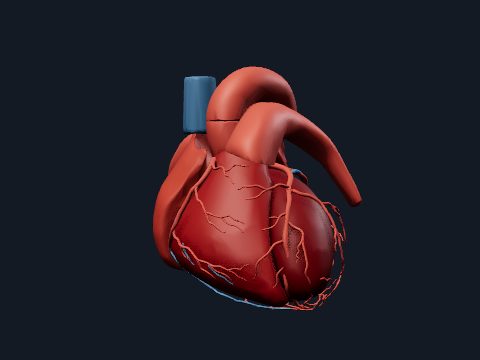
The heart has four valves that keep blood flowing in one direction: the tricuspid and mitral valves between the atria and ventricles, and the pulmonary and aortic valves at the exits of the ventricles. Atrioventricular valves are held by chordae tendineae and papillary muscles; semilunar valves are three pocket-like cusps. In this free 3D model you can study each valve on its own or inside an opened heart.
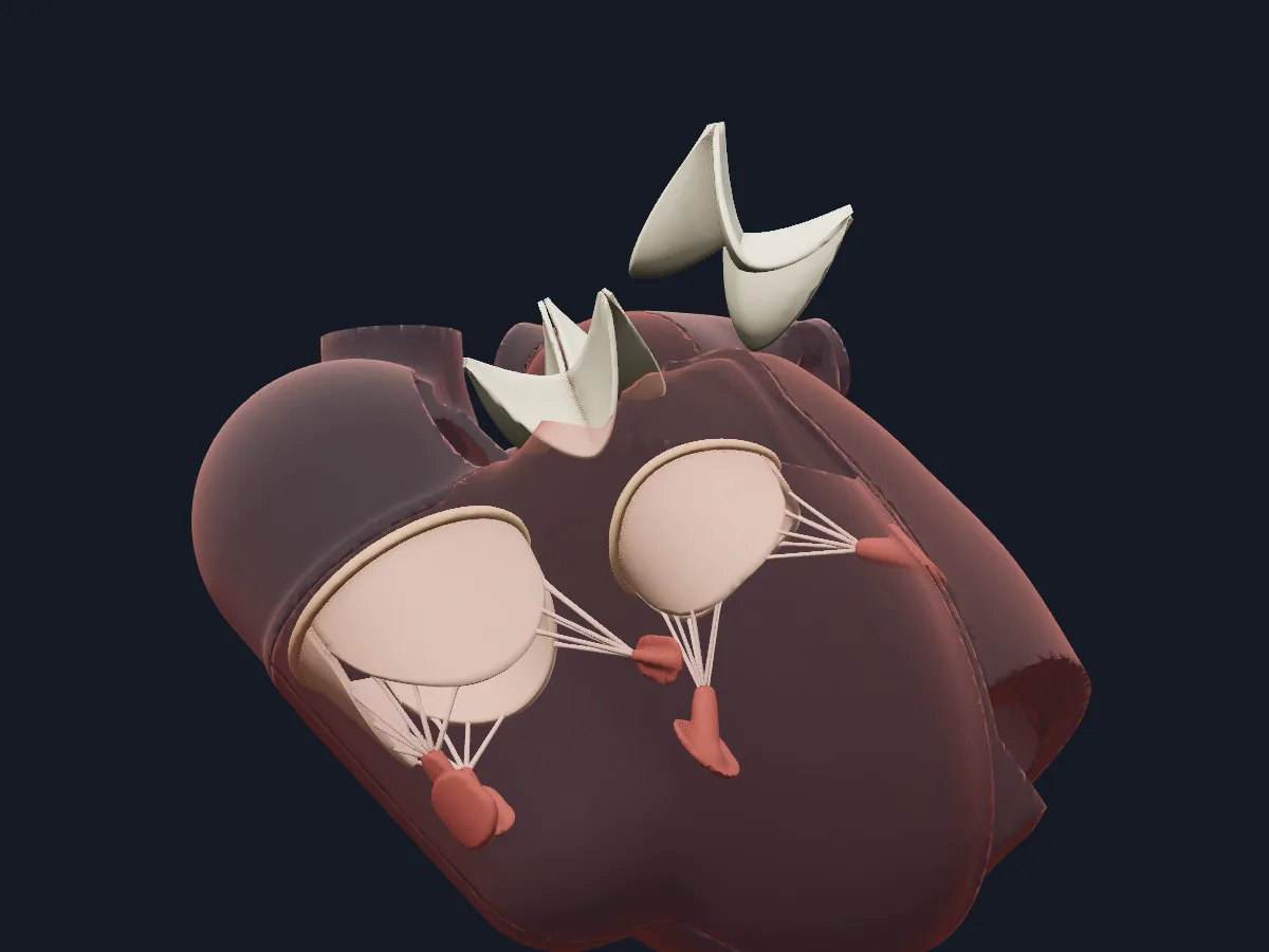 Image: Cogni Anatomy · MIT · Sources: Layered Human
- Tricuspid valve: 3 leaflets (anterior, posterior, septal) and usually 3 papillary muscles
- Mitral valve: 2 leaflets (anterior, posterior) and 2 papillary muscles (anterolateral, posteromedial)
- Aortic valve: 3 cusps (right coronary, left coronary, non-coronary); the coronary arteries arise from the sinuses above the first two
- Pulmonary valve: 3 cusps (anterior, right, left)
- The first heart sound comes from closure of the atrioventricular valves, the second from closure of the semilunar valves
Atrioventricular and semilunar valves
Each atrioventricular valve has leaflets fixed to a fibrous annulus. Thin cords, the chordae tendineae, run from the leaflet edges to papillary muscles on the ventricular wall. When the ventricle contracts, the papillary muscles tighten the chordae so the leaflets close without turning back into the atrium.
The pulmonary and aortic valves have no chordae. Their three cusps fill with returning blood and meet in the centre, closing the outflow. All four valve rings are anchored in the fibrous skeleton of the heart; its central fibrous body lies between the mitral, tricuspid and aortic rings.
What the 3D model shows
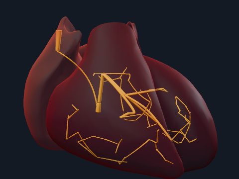
The valves view isolates the valve apparatus in 48 parts: the mitral annulus, both mitral leaflets, the anterolateral and posteromedial papillary muscles with six chordae each; the tricuspid annulus, its anterior, posterior and septal leaflets and three papillary muscles with six chordae each; and the three cusps of both the aortic and pulmonary valves. The open heart view cuts the heart in a coronal plane and adds the walls, septa, central fibrous body, moderator band, coronary vessels and great vessels.
How to study the heart valves
In the valves view, use the superior view to look down on the valve plane: the aortic valve sits in the middle, wedged between the mitral and tricuspid valves, with the pulmonary valve in front. Rotate and follow single chordae from a leaflet to its papillary muscle. Then open the heart view in the anterior view, find the moderator band in the right ventricle and the central fibrous body. Finally, name each valve, its leaflets or cusps and its papillary muscles from memory.
Views in the 3D model
The valve apparatus on its own: annuli, leaflets and cusps, chordae tendineae and papillary muscles of all four valves.
Open this view in 3DImage: Cogni Anatomy · MIT · Sources: Layered Human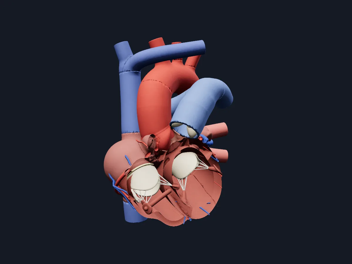
The heart opened in a coronal plane, showing the valves and chordae inside the chambers with the coronary and great vessels.
Open this view in 3DImage: Cogni Anatomy · MIT · Sources: Layered Human
What is in this model
121 3D parts. Names follow the Foundational Model of Anatomy (FMA).
- Heart 69
- Veins 28
- Arteries 24
- Anterior descending artery
- Aortic arch
- Aortic left coronary cusp
- Aortic non coronary cusp
- Aortic right coronary cusp
- Ascending aorta
- Atrioventricular nodal artery
- Atrium anterior wall
- Atrium lateral wall
- Atrium posterior wall
- Auricle
- Brachiocephalic trunk
- Brachiocephalic vein
- Central fibrous body
- Circumflex artery
- Common carotid artery
- Conus artery
- Coronary artery
- Coronary sinus
- Descending aorta
- First anterior cardiac vein
- First diagonal artery
- First obtuse marginal artery
- First septal perforator
- Great cardiac vein
- Great cardiac vein tributary 1
- Great cardiac vein tributary 2
- Great cardiac vein tributary 3
- Great cardiac vein tributary 4
- Inferior pulmonary vein
- Inferior vena cava
- Interatrial septum
- Interventricular septum
- Main coronary artery
- Marginal artery
- Marginal vein
- Marginal vein tributary 1
- Marginal vein tributary 2
- Middle cardiac vein
- Middle cardiac vein tributary 1
- and 73 more
Questions
What is the difference between atrioventricular and semilunar valves?
Atrioventricular valves (tricuspid and mitral) lie between atria and ventricles and are held by chordae tendineae and papillary muscles. Semilunar valves (pulmonary and aortic) guard the outflow of the ventricles and close by the shape of their three cusps alone.
What do the chordae tendineae do?
They connect the leaflets of the tricuspid and mitral valves to the papillary muscles. During ventricular contraction they hold the leaflets in place so the valve closes tightly instead of bulging into the atrium.
Which heart valve has only two leaflets?
The mitral valve, between the left atrium and left ventricle, also called the bicuspid valve. The tricuspid, pulmonary and aortic valves each have three.