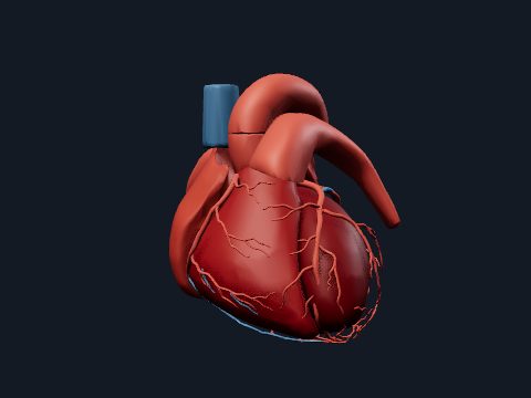
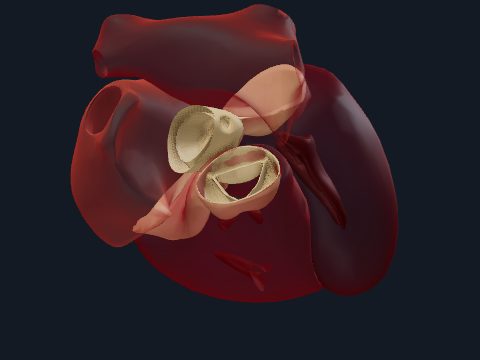
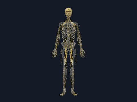
Heart conduction system in 3D
The cardiac conduction system is a pathway of specialised heart muscle cells that starts each heartbeat and spreads it in a fixed order. The sinoatrial node fires, the impulse crosses the atria to the atrioventricular node and then runs through the bundle of His, the bundle branches and the Purkinje fibres into the ventricular walls. In this free 3D model you can see this pathway inside the heart and isolate each of its parts.
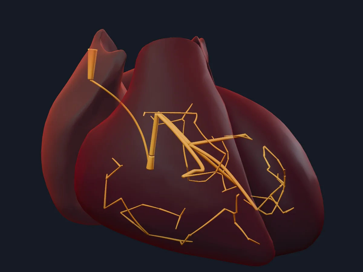 Image: Cogni Anatomy · CC BY 4.0 · Sources: HRA, Cogni
- The sinoatrial node lies in the wall of the right atrium near the opening of the superior vena cava
- The atrioventricular node lies in the interatrial septum, in the triangle of Koch near the opening of the coronary sinus
- The sinoatrial node sets a normal resting rate of about 60–100 beats per minute
- The atrioventricular node delays the impulse by roughly 0.1 s, so the atria finish contracting before the ventricles
- The atrioventricular bundle is normally the only electrical connection between the atria and the ventricles
Parts of the conduction system
The sinoatrial node is the natural pacemaker: its cells depolarise on their own, and autonomic nerves only speed them up or slow them down. The impulse spreads through the atrial muscle along internodal pathways to the atrioventricular node.
The fibrous skeleton insulates the atria from the ventricles, so the impulse must pass through the atrioventricular bundle of His, which pierces the central fibrous body. The bundle divides on the interventricular septum into right and left bundle branches; the right branch reaches the anterior papillary muscle through the moderator band. Purkinje fibres spread beneath the endocardium, activating the ventricles from the apex upward.
What the 3D model shows
The conduction view has 8 parts: the sinoatrial node, the internodal pathways, the atrioventricular node, the atrioventricular bundle, the left and right bundle branches, and the Purkinje fibres of the left and right ventricles. It is a schematic teaching model: the geometry shows where each part lies and how they connect, not the exact microscopic course of the fibres, which cannot be seen with the naked eye in a real heart.
How to study the conduction system
Open the conduction view in the anterior view and trace one impulse in order, clicking each part as you name it: sinoatrial node, internodal pathways, atrioventricular node, bundle of His, bundle branches, Purkinje fibres. Rotate to see where the bundle runs along the septum and how the branches split. Then hide everything and write the sequence from memory, adding for each step where it lies. The open heart view on the heart valves page shows the moderator band that carries the right bundle branch.
What is in this model
8 3D parts. Names follow the Foundational Model of Anatomy (FMA).
- Nerves 8
- Atrioventricular bundle (His)
- Atrioventricular node
- Bundle branch
- Internodal pathways
- Sinoatrial node
- Subendocardial branches (Purkinje fibres) of left ventricle
- Subendocardial branches (Purkinje fibres) of right ventricle
Questions
What is the natural pacemaker of the heart?
The sinoatrial (SA) node in the wall of the right atrium. Its cells generate impulses on their own and set the heart rate.
What is the order of the cardiac conduction system?
Sinoatrial node, atrial muscle and internodal pathways, atrioventricular node, atrioventricular bundle (bundle of His), right and left bundle branches, and Purkinje fibres.
Why is there a delay at the AV node?
Conduction through the atrioventricular node is slow, which gives the atria time to contract and fill the ventricles before the ventricles are activated.