Blood vessels of the trunk in 3D
The blood vessels of the trunk are the aorta with its branches to the chest wall, lungs, gut, kidneys and pelvis, and the veins that return blood through the venae cavae, the azygos system and the hepatic portal vein. In this free 3D model you can follow arteries and veins together or separately against a see-through skeleton and isolate any single branch.
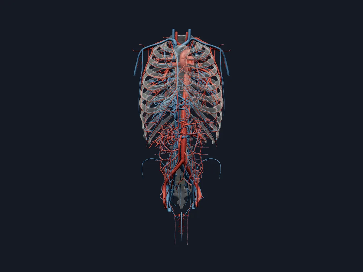 Image: Cogni Anatomy · CC BY 4.0 · Sources: BodyParts3D
- The aorta has 4 parts: ascending aorta, arch, descending thoracic aorta and abdominal aorta
- The arch of the aorta gives 3 branches: brachiocephalic trunk, left common carotid and left subclavian arteries
- The abdominal aorta enters the abdomen at T12 and divides into the common iliac arteries at about L4
- 3 unpaired arteries supply the gut: coeliac trunk (foregut), superior mesenteric (midgut) and inferior mesenteric (hindgut)
- The hepatic portal vein forms behind the neck of the pancreas from the superior mesenteric and splenic veins
Arteries and veins of the trunk
The aorta leaves the left ventricle, arches over the left main bronchus and descends on the left of the vertebral bodies. In the chest it gives posterior intercostal, bronchial and oesophageal branches; in the abdomen, the coeliac trunk, the mesenteric arteries, the renal and suprarenal arteries and paired lumbar arteries.
Venous blood from the head, arms and chest wall drains into the superior vena cava, partly through the azygos and hemiazygos veins. The inferior vena cava runs to the right of the aorta and collects the iliac, lumbar, renal and hepatic veins. Blood from the gut and spleen first passes through the hepatic portal vein to the liver.
What the 3D model shows
The model has about 330 arterial and 275 venous parts. On the arterial side: the aorta in all its parts, the coronary arteries, the pulmonary trunk with segmental pulmonary arteries, the coeliac, hepatic, splenic and pancreaticoduodenal arteries, mesenteric branches down to the sigmoid and superior rectal arteries, segmental renal arteries, and intercostal, internal thoracic and epigastric arteries. On the venous side: both venae cavae, the azygos system, the portal vein with its branches inside the liver, the hepatic, mesenteric, renal and iliac veins, and the cardiac and pulmonary veins.
How to study the trunk vessels
Open the arteries view in the anterior view and follow the aorta from the heart to its bifurcation, naming each branch in order: arch branches, coeliac trunk, superior mesenteric, renal and inferior mesenteric arteries. Isolate the coeliac trunk to see its three branches. Switch to the veins view and turn to the posterior view for the azygos and hemiazygos veins. Then trace the superior mesenteric and splenic veins into the portal vein and on into the liver. Afterwards, draw both trees from memory.
Views in the 3D model
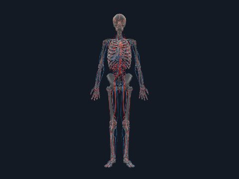
All arteries and veins of the trunk together against a see-through skeleton.
Open this view in 3DImage: Cogni Anatomy · CC BY 4.0 · Sources: BodyParts3D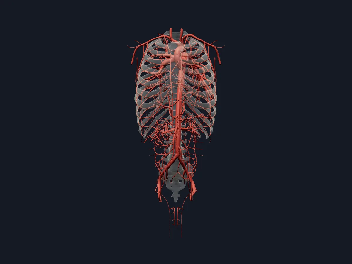
Arteries only, from the aorta and coronary arteries to the mesenteric, renal and intercostal branches.
Open this view in 3DImage: Cogni Anatomy · CC BY 4.0 · Sources: BodyParts3D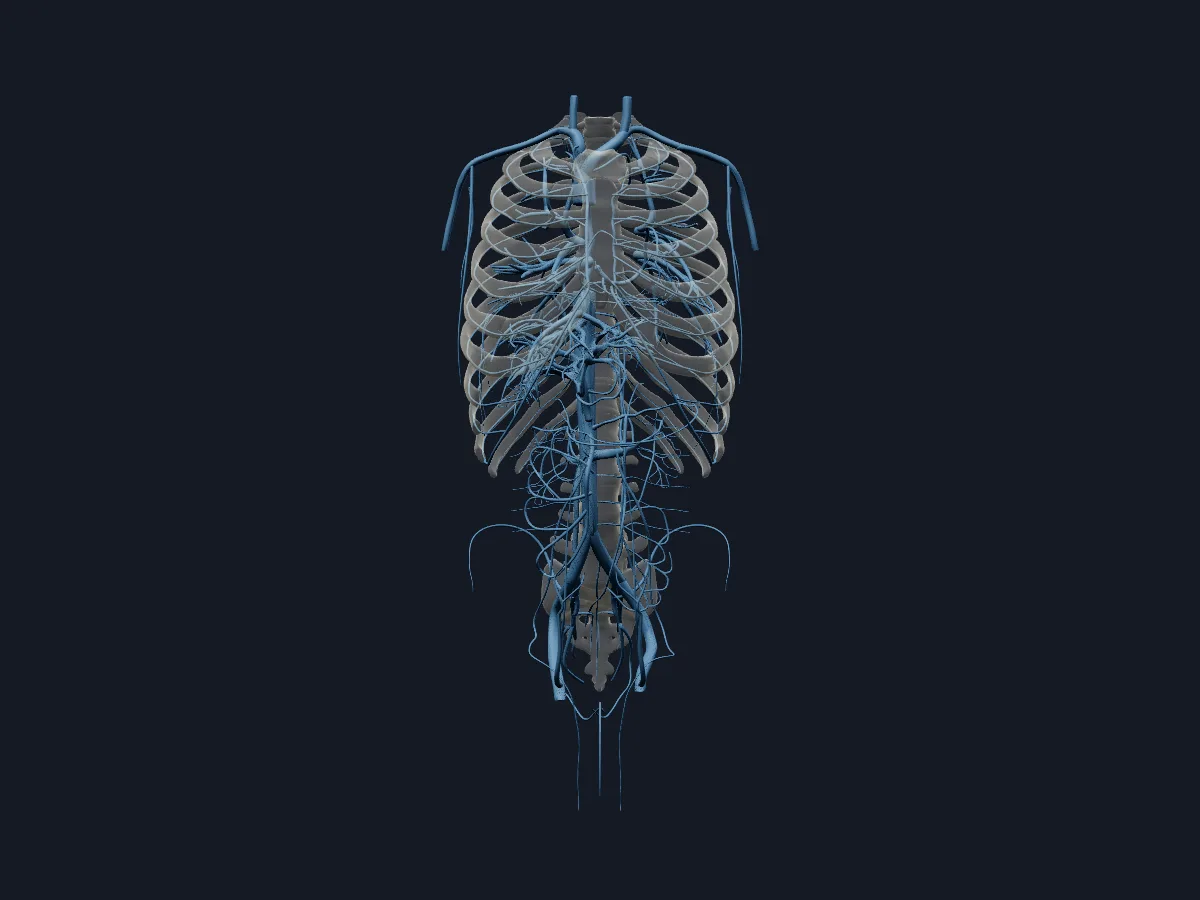
Veins only, including both venae cavae, the azygos system and the hepatic portal vein.
Open this view in 3DImage: Cogni Anatomy · CC BY 4.0 · Sources: BodyParts3D
What is in this model
618 3D parts. Names follow the Foundational Model of Anatomy (FMA).
- Arteries 339
- Veins 279
- Abdominal aorta
- Accessory hemiazygos vein
- Accessory subsuperior branch of right posterior basal segmental artery
- Acromial branch of left thoraco-acromial artery
- Acromial branch of right thoraco-acromial artery
- Anterior basal segmental artery
- Anterior basal segmental vein
- Anterior branch of lateral segmental artery
- Anterior branch of right anterior segmental artery
- Anterior cardiac vein
- Anterior cecal artery
- Anterior division of left renal artery
- Anterior division of right renal artery
- Anterior inferior pancreaticoduodenal artery
- Anterior inferior segmental branch of right portal vein
- Anterior inferior segmental hepatic artery
- Anterior inferior segmental tributary of middle hepatic vein
- Anterior interventricular vein
- Anterior part of lateral segmental vein
- Anterior part of right apical segmental artery
- Anterior part of right apical segmental vein
- Anterior segmental artery
- Anterior segmental vein
- Anterior superior pancreaticoduodenal artery
- Anterior superior segmental branch of right portal vein
- Anterior superior segmental hepatic artery
- Anterior superior segmental tributary of right hepatic vein
- Apical part of right apical segmental artery
- Apical part of right apical segmental vein
- Apical part of right posterior segmental artery
- Apical segmental artery
- Apical segmental vein
- Appendicular artery
- Arch of aorta
- Ascending aorta
- Ascending branch of inferior branch of ileocolic artery
- Ascending branch of left colic artery
- Ascending lumbar vein
- Axillary artery
- Axillary vein
- and 220 more
Questions
What are the branches of the abdominal aorta?
Unpaired anterior branches to the gut (coeliac trunk, superior and inferior mesenteric arteries), paired branches to organs (suprarenal, renal and gonadal arteries), paired parietal branches (inferior phrenic and lumbar arteries) and the median sacral artery, before it divides into the common iliac arteries.
What is the difference between the portal vein and the hepatic veins?
The hepatic portal vein brings blood from the gut, pancreas and spleen into the liver. The hepatic veins drain blood out of the liver into the inferior vena cava.
What does the azygos vein drain?
The posterior chest wall through the posterior intercostal veins, together with bronchial and oesophageal veins. It arches over the right main bronchus and ends in the superior vena cava.