Nervous system in 3D
The nervous system controls and coordinates the body. Its central part is the brain and spinal cord; its peripheral part consists of 12 pairs of cranial nerves, 31 pairs of spinal nerves with their plexuses and branches, and the autonomic nerves and ganglia. In this free 3D model you can follow the nerves through the whole body over a see-through skeleton, show the muscles they run between and isolate any nerve by name.
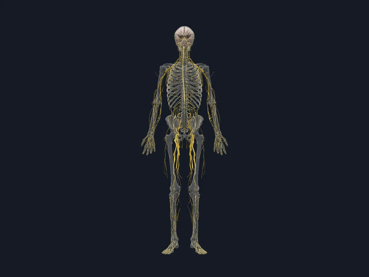 Image: Cogni Anatomy · CC BY-SA 4.0 · Sources: BodyParts3D, HRA, Z-Anatomy
- Central nervous system: brain and spinal cord
- Peripheral nervous system: 12 pairs of cranial nerves and 31 pairs of spinal nerves
- The brachial plexus is formed by the anterior rami of C5–T1, the lumbar plexus by L1–L4
- The sciatic nerve (L4–S3) is the largest nerve in the body
- Sympathetic fibres leave the spinal cord only from segments T1–L2
Central, peripheral and autonomic parts
Each spinal nerve forms from an anterior (motor) root and a posterior (sensory) root with its spinal ganglion, then divides into anterior and posterior rami. The anterior rami form the cervical, brachial, lumbar and sacral plexuses, which supply the neck and limbs, while in the thorax they continue as the intercostal nerves.
The autonomic nervous system controls the viscera, vessels and glands. Sympathetic outflow arises from the thoracic and upper lumbar spinal cord. Sympathetic preganglionic fibres synapse in paravertebral or prevertebral ganglia; fibres to the adrenal medulla form a special pathway. Its parasympathetic part travels with cranial nerves III, VII, IX and X and the sacral nerves S2–S4.
What the 3D model shows
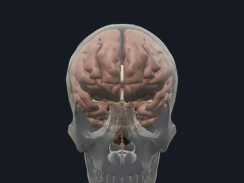
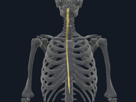
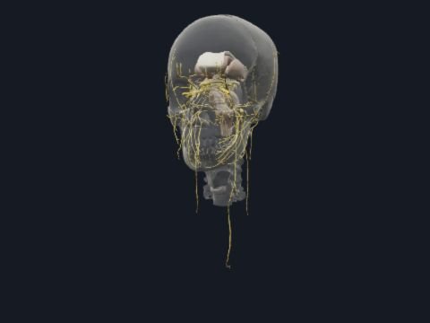
The nerves view has 423 parts over a faded skeleton: the brain, the spinal cord segments C1–S5 and the cauda equina, the cranial nerves, spinal nerve roots and a spinal ganglion, and the sympathetic trunk with its ganglia. In the upper limb it follows the brachial plexus through its roots, trunks and divisions into the axillary, musculocutaneous, median, ulnar and radial nerves down to the digital branches. The trunk has the intercostal, iliohypogastric, ilio-inguinal and genitofemoral nerves; the lower limb has the femoral, obturator, sciatic, tibial and fibular nerves, the plantar nerves and the pudendal nerve. The second view adds about 500 muscle parts.
How to study the nervous system
In the nerves view, pick one limb nerve and trace it from its spinal cord segments to its last branch: for example, the median nerve from the brachial plexus through the arm and forearm to the fingers. From the posterior view, follow the sciatic nerve down the thigh until it divides into the tibial and common fibular nerves. Then switch to the nerves and muscles view to see which muscles each nerve runs between. Close the model and list the terminal branches of the brachial plexus and the nerves of the lower limb from memory.
Views in the 3D model
The nerves of the whole body, from the brain and spinal cord to the limb branches, over a faded skeleton.
Open this view in 3DImage: Cogni Anatomy · CC BY-SA 4.0 · Sources: BodyParts3D, HRA, Z-Anatomy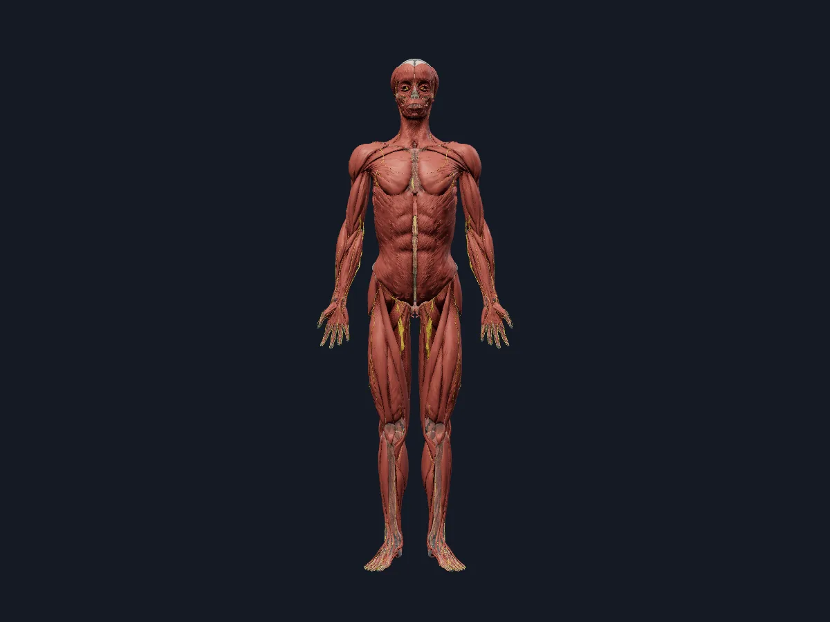
The same nerves shown with the muscles of the whole body.
Open this view in 3DImage: Cogni Anatomy · CC BY-SA 4.0 · Sources: BodyParts3D, HRA, Z-Anatomy
What is in this model
921 3D parts. Names follow the Foundational Model of Anatomy (FMA).
- Muscles 498
- Nerves 423
- (Communicating branch of median nerve with ulnar nerve)
- Abdominal part of left pectoralis major
- Abdominal part of right pectoralis major
- Abducens nerve (VI)
- Abductor digiti minimi of left foot
- Abductor digiti minimi of left hand
- Abductor digiti minimi of right foot
- Abductor digiti minimi of right hand
- Abductor hallucis
- Abductor pollicis brevis
- Abductor pollicis longus
- Accessory nerve (XI)
- Acromial part of left deltoid
- Acromial part of right deltoid
- Adductor brevis
- Adductor longus
- Adductor magnus
- Adductor minimus
- Amygdala
- Anconeus
- Angular gyrus
- Anterior belly of digastric muscle
- Anterior branch of medial antebrachial cutaneous nerve
- Anterior branch of obturator nerve
- Anterior commissure
- Anterior cutaneous branches of femoral nerve
- Anterior division of inferior trunk of brachial plexus
- Anterior division of mandibular nerve
- Anterior division of middle trunk of brachial plexus
- Anterior division of superior trunk of brachial plexus
- Anterior ethmoidal nerve
- Anterior interosseous nerve of forearm
- Anterior papillary muscle of right ventricle
- Anterior part of left superior temporal gyrus
- Anterior part of right superior temporal gyrus
- Anterior root of posterior femoral cutaneous nerve
- Anterior root of spinal nerve
- Anterolateral head of lateral papillary muscle of left ventricle
- Ary-epiglottic part of oblique arytenoid muscle
- Aryepiglotticus
- and 497 more
Questions
What are the two main parts of the nervous system?
The central nervous system, made up of the brain and spinal cord, and the peripheral nervous system, made up of the cranial and spinal nerves, their branches and ganglia.
How many pairs of spinal nerves are there?
31 pairs: 8 cervical, 12 thoracic, 5 lumbar, 5 sacral and 1 coccygeal.
What is the largest nerve in the human body?
The sciatic nerve, formed from the L4–S3 roots of the sacral plexus. It runs down the back of the thigh and usually divides above the knee into the tibial and common fibular nerves.